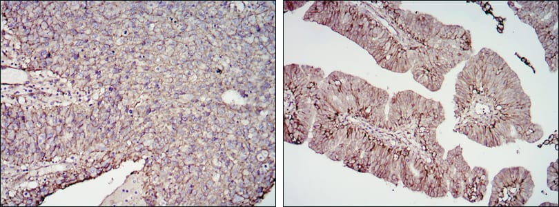
CD276 Primary Antibody

Item Information
Catalog #
Size
Price
Description

Costimulatory B7 molecules (e.g., B7-1, or CD80; MIM 112203) signal through CD28 (MIM 186760) family molecules such as CD28, CTLA4 (MIM 123890), and ICOS (MIM 604558). May participate in the regulation of T-cell-mediated immune response. May play a protective role in tumor cells by inhibiting natural-killer mediated cell lysis as well as a role of marker for detection of neuroblastoma cells. May be involved in the development of acute and chronic transplant rejection and in the regulation of lymphocytic activity at mucosal surfaces. Could also play a key role in providing the placenta and fetus with a suitable immunological environment throughout pregnancy. Both isoform 1 and isoform 2 appear to be redundant in their ability to modulate CD4 T-cell responses. Isoform 2 is shown to enhance the induction of cytotoxic T-cells and selectively stimulates interferon gamma production in the presence of T-cell receptor signaling.

Product Overview
Entrez GenelD
80381
Aliases
B7H3; B7-H3; 4Ig-B7-H3; CD276
Clone#
6A1
Host / Isotype
Mouse / IgG1
Species Reactivity
Human
Immunogen
Purified recombinant fragment of human CD276 expressed in E. Coli.
Formulation
Ascitic fluid containing 0.03% sodium azide.
Storage
4°C; -20°C for long term storage
Product Applications
WB (Western Blot)
1/500 - 1/2000
IHC_P(Immunohistochemistry)
1/200 - 1/1000
FCM (Flow Cytometry)
1/200 - 1/400
ELISA
1/10000
References
1. Genome Res. 2004 Oct;14(10B):2121-7.
2. Cell Mol Immunol. 2005 Aug;2(4):307-11.
Product Image
Western Blot
Figure 1: Western blot analysis using CD276 mAb against HEK293 (1) and CD276(AA: 30-130)-hIgGFc transfected HEK293 (2) cell lysate.
Immunohistochemical analysis
Figure 2: Immunohistochemical analysis of paraffin-embedded cervical cancer tissues (left) and ovarian cancer tissues (right) using CD276 mouse mAb with DAB staining.
Flow cytometric
Figure 3: Flow cytometric analysis of PC-3 cells using CD276 mouse mAb (green) and negative control (purple).
For Research Use Only. Not for use in diagnostic procedures.