

CD267 Primary Antibody
Item Information
Catalog #
Size
Price
Description
The protein encoded by this gene is a lymphocyte-specific member of the tumor necrosis factor (TNF) receptor superfamily. It interacts with calcium-modulator and cyclophilin ligand (CAML). The protein induces activation of the transcription factors NFAT, AP1, and NF-kappa-B and plays a crucial role in humoral immunity by interacting with a TNF ligand. This gene is located within the Smith-Magenis syndrome region on chromosome 17. [provided by RefSeq, Jul 2008]
Product Overview
Entrez GenelD
23495
Aliases
CVID; RYZN; TACI; CD267; CVID2; IGAD2; TNFRSF14B
Clone#
1F6B3
Host / Isotype
Mouse / Mouse IgG1
Immunogen
Purified recombinant fragment of human CD267 (AA: extra(1-165)) expressed in E. Coli.
Formulation
Purified antibody in PBS with 0.05% sodium azide
Storage
Store at 4°C short term. Aliquot and store at -20°C long term. Avoid freeze/thaw cycles.
Product Applications
WB (Western Blot)
1/500 - 1/2000
IHC_P(Immunohistochemistry)
1/200-1/1000
FCM (Flow Cytometry)
1/200-1/400
ELISA
1/10000
References
1,Front Immunol. 2018 Oct 2;9:2125.2,J Thromb Haemost. 2017 Nov;15(11):2259-2269.
Product Image
ELISA

Figure 1: Black line: Control Antigen (100 ng);Purple line: Antigen (10ng); Blue line: Antigen (50 ng); Red line: Antigen (100 ng)
WESTERN BLOT

Figure 2: Western blot analysis using CD267 mAb against human CD267 (AA: extra(1-165)) recombinant protein. (Expected MW is 44.5kDa)
WESTERN BLOT

Figure 3: Western blot analysis using CD267 mAb against HEK293-6e (1) and CD267 (AA: extra(1-165))-hIgGFc transfected HEK293-6e (2) cell lysate.
FLOW CYTOMETRY
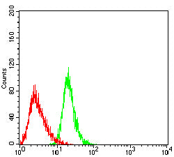
Figure 4: Flow cytometric analysis of Jurkat cells using CD267 mouse mAb (green) and negative control (red).
FLOW CYTOMETRY

Figure 5: Flow cytometric analysis of THP-1 cells using CD267 mouse mAb (green) and negative control (red).
IMMUNOHISTOCHEMISTRY

Figure 6: Immunohistochemical analysis of paraffin-embedded cervical cancer tissues using CD267 mouse mAb with DAB staining.
IMMUNOHISTOCHEMISTRY

Figure 7: Immunohistochemical analysis of paraffin-embedded lymphoid tissues using CD267 mouse mAb with DAB staining.
For Research Use Only. Not for use in diagnostic procedures.