
CD247 Primary Antibody
Item Information
Catalog #
Size
Price
Description
The protein encoded by this gene is T-cell receptor zeta, which together with T-cell receptor alpha/beta and gamma/delta heterodimers, and with CD3-gamma, -delta and -epsilon, forms the T-cell receptor-CD3 complex. The zeta chain plays an important role in coupling antigen recognition to several intracellular signal-transduction pathways. Low expression of the antigen results in impaired immune response. Two alternatively spliced transcript variants encoding distinct isoforms have been found for this gene.
Product Overview
Entrez GenelD
919
Aliases
T3Z; CD3H; CD3Q; CD3Z; TCRZ; CD3-ZETA; CD247
Clone#
4B10
Host / Isotype
Mouse / IgG1
Species Reactivity
Human
Immunogen
Purified recombinant fragment of human CD247 expressed in E. Coli.
Formulation
Ascitic fluid containing 0.03% sodium azide.
Storage
Store at 4°C short term. Aliquot and store at -20°C long term. Avoid freeze/thaw cycles.
Product Applications
WB (Western Blot)
1/500 - 1/2000
IHC_P(Immunohistochemistry)
1/200 - 1/1000
ICC (Immunocytochemistry)
1/200 - 1/1000
FCM (Flow Cytometry)
1/200 - 1/400
ELISA
1/10000
References
1. J Immunol. 2002 Aug 15;169(4):1705-12.
2. Arthritis Rheum. 2003 Jul;48(7):1948-55.
3. Nat Methods. 2005 Aug;2(8):591-8.
2. Arthritis Rheum. 2003 Jul;48(7):1948-55.
3. Nat Methods. 2005 Aug;2(8):591-8.
Product Image
Western Blot

Figure 1: Western blot analysis using CD247 mAb against CD247(AA: 52-164)-hIgGFc transfected HEK293 cell lysate.
Immunohistochemical analysis

Figure 2: Immunohistochemical analysis of paraffin-embedded human Thymus tissues using anti-CD247 mouse mAb
Immunofluorescence analysis
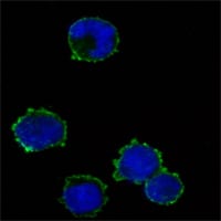
Figure3: Immunofluorescence analysis of K562 cells using anti-CD247 mAb (green). Blue: DRAQ5 fluorescent DNA dye.
Flow cytometric
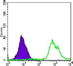
Figure 4: Flow cytometric analysis of Jurkat cells using anti-CD247 mAb (green) and negative control (purple).
For Research Use Only. Not for use in diagnostic procedures.