

CD239 Primary Antibody
Item Information
Catalog #
Size
Price
Description
This gene encodes Lutheran blood group glycoprotein, a member of the immunoglobulin superfamily and a receptor for the extracellular matrix protein, laminin. The protein contains five extracellular immunoglobulin domains, a single transmembrane domain, and a short C-terminal cytoplasmic tail. This protein may play a role in epithelial cell cancer and in vaso-occlusion of red blood cells in sickle cell disease. Polymorphisms in this gene define some of the antigens in the Lutheran system and also the Auberger system. Inactivating variants of this gene result in the recessive Lutheran null phenotype, Lu(a-b-), of the Lutheran blood group. Two transcript variants encoding different isoforms have been found for this gene.
Product Overview
Entrez GenelD
67.4kDa
Aliases
BCAM; AU; LU; MSK19
Clone#
5C5A6
Host / Isotype
Mouse / IgG2a
Species Reactivity
Human
Immunogen
Purified recombinant fragment of human CD239 (AA: extra 32-197) expressed in E. Coli.
Formulation
Purified antibody in PBS with 0.05% sodium azide
Storage
Store at 4°C short term. Aliquot and store at -20°C long term. Avoid freeze/thaw cycles.
Product Applications
WB (Western Blot)
1/500 - 1/2000
IHC_P(Immunohistochemistry)
1/200 - 1/1000
FCM (Flow Cytometry)
1/200 - 1/400
ELISA
1/10000
References
1.J Biomed Sci. 2017 Aug 26;24(1):61.
2.Exp Cell Res. 2014 Oct 15;328(1):197-206.
2.Exp Cell Res. 2014 Oct 15;328(1):197-206.
Product Image
Elisa

Figure 1:Black line: Control Antigen (100 ng);Purple line: Antigen (10ng); Blue line: Antigen (50 ng); Red line:Antigen (100 ng)
Western Blot
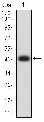
Figure 2:Western blot analysis using CD239 mAb against human CD239 (AA: extra 32-197) recombinant protein. (Expected MW is 44.1 kDa)
Western Blot

Figure 3:Western blot analysis using CD239 mAb against HEK293 (1) and CD239 (AA: extra 32-197)-hIgGFc transfected HEK293 (2) cell lysate.
Flow cytometric

Figure 4:Flow cytometric analysis of MOLT4 cells using CD239 mouse mAb (green) and negative control (red).
Immunohistochemical analysis

Figure 5:Immunohistochemical analysis of paraffin-embedded lung cancer tissues using CD239 mouse mAb with DAB staining.
Immunohistochemical analysis

Figure 6:Immunohistochemical analysis of paraffin-embedded ovarian cancer tissues using CD239 mouse mAb with DAB staining.
For Research Use Only. Not for use in diagnostic procedures.