

CD235a Primary Antibody
Item Information
Catalog #
Size
Price
Description
Glycophorins A (GYPA) and B (GYPB) are major sialoglycoproteins of the human erythrocyte membrane which bear the antigenic determinants for the MN and Ss blood groups. In addition to the M or N and S or s antigens that commonly occur in all populations, about 40 related variant phenotypes have been identified. These variants include all the variants of the Miltenberger complex and several isoforms of Sta, as well as Dantu, Sat, He, Mg, and deletion variants Ena, S-s-U- and Mk. Most of the variants are the result of gene recombinations between GYPA and GYPB.
Product Overview
Entrez GenelD
2993
Aliases
MN; GPA; MNS; GPSAT; PAS-2; GPErik; HGpMiV; HGpMiXI; HGpSta(C)
Clone#
2D5A6
Host / Isotype
Mouse / Mouse IgG1
Species Reactivity
Human
Immunogen
Purified recombinant fragment of human CD235a (AA: 20-91) expressed in E. Coli.
Formulation
Purified antibody in PBS with 0.05% sodium azide
Storage
Store at 4°C short term. Aliquot and store at -20°C long term. Avoid freeze/thaw cycles.
Product Applications
WB (Western Blot)
1/500 - 1/2000
FCM (Flow Cytometry)
1/200 - 1/400
ELISA
1/10000
References
1.Transfusion. 2020 Jun;60(6):1287-1293.
2.Hum Genet. 2018 Feb;137(2):151-160.
2.Hum Genet. 2018 Feb;137(2):151-160.
Product Image
Elisa

Figure 1:Black line: Control Antigen (100 ng);Purple line: Antigen (10ng); Blue line: Antigen (50 ng); Red line:Antigen (100 ng)
Western Blot
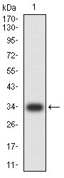
Figure 2:Western blot analysis using CD235a mAb against human CD235a (AA: 20-91) recombinant protein. (Expected MW is 34 kDa)
Western Blot
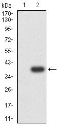
Figure 3:Western blot analysis using CD235a mAb against HEK293-6e (1) and CD235a (AA: 20-91)-hIgGFc transfected HEK293-6e (2) cell lysate.
Immunofluorescence analysis

Figure 4:Flow cytometric analysis of K562 cells using CD235a mouse mAb (green) and negative control (red).
For Research Use Only. Not for use in diagnostic procedures.