
CD227 Primary Antibody
Item Information
Catalog #
Size
Price
Description
This gene encodes a membrane-bound protein that is a member of the mucin family. Mucins are O-glycosylated proteins that play an essential role in forming protective mucous barriers on epithelial surfaces. These proteins also play a role in intracellular signaling. This protein is expressed on the apical surface of epithelial cells that line the mucosal surfaces of many different tissues including lung, breast stomach and pancreas. This protein is proteolytically cleaved into alpha and beta subunits that form a heterodimeric complex. The N-terminal alpha subunit functions in cell-adhesion and the C-terminal beta subunit is involved in cell signaling. Overexpression, aberrant intracellular localization, and changes in glycosylation of this protein have been associated with carcinomas. This gene is known to contain a highly polymorphic variable number tandem repeats (VNTR) domain. Alternate splicing results in multiple transcript variants.
Product Overview
Entrez GenelD
4582
Aliases
MUC1; EMA; MCD; PEM; PUM; KL-6; MAM6; MCKD; PEMT; H23AG; MCKD1; MUC-1; ADMCKD; ADMCKD1; CA 15-3; MUC-1/X; MUC1/ZD; MUC-1/SEC
Clone#
3B10B4
Host / Isotype
Mouse / IgG1
Species Reactivity
Human
Immunogen
Purified recombinant fragment of human CD227 (AA: extra 66-175) expressed in E. Coli.
Formulation
Purified antibody in PBS with 0.05% sodium azide
Storage
Store at 4°C short term. Aliquot and store at -20°C long term. Avoid freeze/thaw cycles.
Product Applications
WB (Western Blot)
1/500 - 1/2000
IHC_P(Immunohistochemistry)
1/200 - 1/1000
FCM (Flow Cytometry)
1/200 - 1/400
ELISA
1/10000
References
1.Pol J Pathol. 2016;67(4):384-391.
2.Blood. 2015 Jul 16;126(3):354-62.
2.Blood. 2015 Jul 16;126(3):354-62.
Product Image
Elisa

Figure 1: Black line: Control Antigen (100 ng);Purple line: Antigen (10ng); Blue line: Antigen (50 ng); Red line:Antigen (100 ng)
Western Blot

Figure 2:Western blot analysis using CD227 mAb against human CD227 (AA: extra 66-175) recombinant protein. (Expected MW is 38.5 kDa)
Western Blot

Figure 3:Western blot analysis using CD227 mAb against HEK293 (1) and CD227 (AA: extra 66-175)-hIgGFc transfected HEK293 (2) cell lysate.
Flow cytometric

Figure 4:Flow cytometric analysis of Raji cells using CD227 mouse mAb (green) and negative control (red).
Immunohistochemical analysis
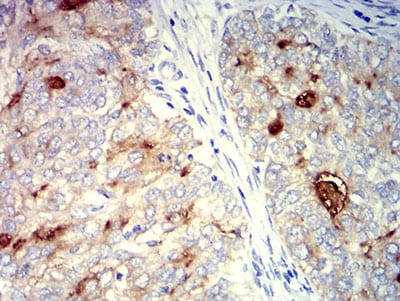
Figure 5:Immunohistochemical analysis of paraffin-embedded cervical cancer tissues using CD227 mouse mAb with DAB staining.
Immunohistochemical analysis

Figure 6:Immunohistochemical analysis of paraffin-embedded breast cancer tissues using CD227 mouse mAb with DAB staining.
For Research Use Only. Not for use in diagnostic procedures.