
CD22 Primary Antibody
Item Information
Catalog #
Size
Price
Description
CD22 may be involved in the localization of B-cells in lymphoid tissues. Binds sialylated glycoproteins; one of which is CD45. Preferentially binds to alpha-2,6-linked sialic acid. The sialic acid recognition site can be masked by cis interactions with sialic acids on the same cell surface. Upon ligand induced tyrosine phosphorylation in the immune response seems to be involved in regulation of B-cell antigen receptor signaling. Plays a role in positive regulation through interaction with Src family tyrosine kinases and may also act as an inhibitory receptor by recruiting cytoplasmic phosphatases via their SH2 domains that block signal transduction through dephosphorylation of signaling molecules
Product Overview
Entrez GenelD
933
Aliases
SIGLEC2; SIGLEC-2
Clone#
1A3A11
Host / Isotype
Mouse / IgG1
Species Reactivity
Human
Immunogen
Purified recombinant fragment of human CD22 (AA: 621-725) expressed in E. Coli.
Formulation
Purified antibody in PBS with 0.05% sodium azide
Storage
Store at 4°C short term. Aliquot and store at -20°C long term. Avoid freeze/thaw cycles.
Product Applications
WB (Western Blot)
1/500 - 1/2000
ICC (Immunocytochemistry)
1/200 - 1/1000
FCM (Flow Cytometry)
1/200 - 1/400
ELISA
1/10000
References
1. Cancer Res. 2012 Nov 1;72(21):5556-65.
2. J Innate Immun. 2011;3(4):411-9.
2. J Innate Immun. 2011;3(4):411-9.
Product Image
Western Blot

Figure 1: Western blot analysis using CD22 mAb against human CD22 recombinant protein. (Expected MW is 37 kDa)
Western Blot

Figure 2: Western blot analysis using CD22 mAb against HEK293 (1) and CD22 (AA: 621-725)-hIgGFc transfected HEK293 (2) cell lysate.
Immunofluorescence analysis

Figure 3: Immunofluorescence analysis of Hela cells using CD22 mouse mAb (green). Blue: DRAQ5 fluorescent DNA dye. Red: Actin filaments have been labeled with Alexa Fluor-555 phalloidin.
Flow cytometric

Figure 4: Flow cytometric analysis of Hela cells using CD22 mouse mAb (green) and negative control (red).
Elisa
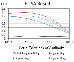
Black line: Control Antigen (100 ng); Purple line: Antigen(10ng); Blue line: Antigen (50 ng); Red line: Antigen (100 ng);
For Research Use Only. Not for use in diagnostic procedures.