

CD21 Primary Antibody
Item Information
Catalog #
Size
Price
Description
This gene encodes a membrane protein, which functions as a receptor for Epstein-Barr virus (EBV) binding on B and T lymphocytes. Genetic variations in this gene are associated with susceptibility to systemic lupus erythematosus type 9 (SLEB9). Alternatively spliced transcript variants encoding different isoforms have been found for this gene.
Product Overview
Entrez GenelD
1380
Aliases
CR2; CR; C3DR; CVID7; SLEB9
Clone#
2D2H6
Host / Isotype
Mouse / Mouse IgG1
Immunogen
Purified recombinant fragment of human CD21 (AA: extra 740-964) expressed in E. Coli.
Formulation
Purified antibody in PBS with 0.05% sodium azide
Storage
Store at 4°C short term. Aliquot and store at -20°C long term. Avoid freeze/thaw cycles.
Product Applications
WB (Western Blot)
1/500 - 1/2000
FCM (Flow Cytometry)
1/200-1/400
ELISA
1/10000
References
1.J Allergy Clin Immunol Pract. 2017 Nov-Dec;5(6):1765-1767.e3. 2.Blood. 2016 Oct 6;128(14):1789-1799.
Product Image
ELISA
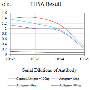
Figure 1: Black line: Control Antigen (100 ng);Purple line: Antigen (10ng); Blue line: Antigen (50 ng); Red line: Antigen (100 ng)
WESTERN BLOT
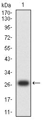
Figure 2: Western blot analysis using CD21 mAb against human CD21 (AA: extra 740-964) recombinant protein. (Expected MW is 27.6 kDa)
WESTERN BLOT
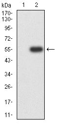
Figure 3: Western blot analysis using CD21 mAb against HEK293-6e (1) and CD21 (AA: extra 740-964)-hIgGFc transfected HEK293 (2) cell lysate.
FLOW CYTOMETRY
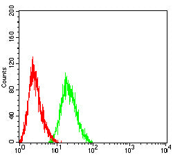
Figure 4: Flow cytometric analysis of HL-60 cells using CD21 mouse mAb (green) and negative control (red).
FLOW CYTOMETRY

Figure 5: Flow cytometric analysis of Jurkat cells using CD21 mouse mAb (green) and negative control (red).
For Research Use Only. Not for use in diagnostic procedures.