

CD209 Primary Antibody
Item Information
Catalog #
Size
Price
Description
This gene encodes a transmembrane receptor and is often referred to as DC-SIGN because of its expression on the surface of dendritic cells and macrophages. The encoded protein is involved in the innate immune system and recognizes numerous evolutionarily divergent pathogens ranging from parasites to viruses with a large impact on public health. The protein is organized into three distinct domains: an N-terminal transmembrane domain, a tandem-repeat neck domain and C-type lectin carbohydrate recognition domain. The extracellular region consisting of the C-type lectin and neck domains has a dual function as a pathogen recognition receptor and a cell adhesion receptor by binding carbohydrate ligands on the surface of microbes and endogenous cells. The neck region is important for homo-oligomerization which allows the receptor to bind multivalent ligands with high avidity. Variations in the number of 23 amino acid repeats in the neck domain of this protein are rare but have a significant impact on ligand binding ability. This gene is closely related in terms of both sequence and function to a neighboring gene (GeneID 10332; often referred to as L-SIGN). DC-SIGN and L-SIGN differ in their ligand-binding properties and distribution. Alternative splicing results in multiple variants.
Product Overview
Entrez GenelD
30835
Aliases
CDSIGN; CLEC4L; DC-SIGN; DC-SIGN1
Clone#
5C2A6
Host / Isotype
Mouse / IgG1
Species Reactivity
Human
Immunogen
Purified recombinant fragment of human CD209 (AA: extra 270-404) expressed in E. Coli.
Formulation
Purified antibody in PBS with 0.05% sodium azide
Storage
Store at 4°C short term. Aliquot and store at -20°C long term. Avoid freeze/thaw cycles.
Product Applications
WB (Western Blot)
1/500 - 1/2000
FCM (Flow Cytometry)
1/200 - 1/400
ELISA
1/10000
References
1.Blood. 2015 Oct 15;126(16):1911-20.
2.PLoS One. 2014 Aug 22;9(8):e105236.
2.PLoS One. 2014 Aug 22;9(8):e105236.
Product Image
Elisa
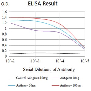
Figure 1: Black line: Control Antigen (100 ng);Purple line: Antigen (10ng); Blue line: Antigen (50 ng); Red line:Antigen (100 ng)
Western Blot
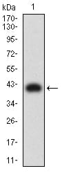
Figure 2:Western blot analysis using CD209 mAb against human CD209 (AA: extra 270-404) recombinant protein. (Expected MW is 41.1 kDa)
Western Blot
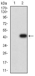
Figure 3:Western blot analysis using CD209 mAb against HEK293 (1) and CD209 (AA: extra 270-404)-hIgGFc transfected HEK293 (2) cell lysate.
Western Blot
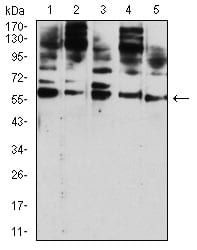
Figure 4:Western blot analysis using CD209 mouse mAb against Hela (1), U937 (2), THP-1 (3), HL-60 (4), and A431 (5) cell lysate.
Flow cytometric
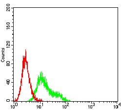
Figure 5:Flow cytometric analysis of Ramos cells using CD209 mouse mAb (green) and negative control (red).
For Research Use Only. Not for use in diagnostic procedures.