

CD203C Primary Antibody
Item Information
Catalog #
Size
Price
Description
The protein encoded by this gene belongs to a series of ectoenzymes that are involved in hydrolysis of extracellular nucleotides. These ectoenzymes possess ATPase and ATP pyrophosphatase activities and are type II transmembrane proteins. Expression of the related rat mRNA has been found in a subset of immature glial cells and in the alimentary tract. The corresponding rat protein has been detected in the pancreas, small intestine, colon, and liver. The human mRNA is expressed in glioma cells, prostate, and uterus. Expression of the human protein has been detected in uterus, basophils, and mast cells. Two transcript variants, one protein coding and the other non-protein coding, have been found for this gene.
Product Overview
Entrez GenelD
5169
Aliases
ENPP3; B10; NPP3; PDNP3; PD-IBETA
Clone#
4C1H2
Host / Isotype
Mouse / IgG1
Species Reactivity
Human
Immunogen
Purified recombinant fragment of human CD203C (AA: extra 45-163) expressed in E. Coli.
Formulation
Purified antibody in PBS with 0.05% sodium azide
Storage
Store at 4°C short term. Aliquot and store at -20°C long term. Avoid freeze/thaw cycles.
Product Applications
WB (Western Blot)
1/500 - 1/2000
IHC_P(Immunohistochemistry)
1/200 - 1/1000
FCM (Flow Cytometry)
1/200 - 1/400
ELISA
1/10000
References
1.Leuk Lymphoma. 2014 Jan;55(1):92-6. 2.J Allergy Clin Immunol. 2010 Feb;125(2):483-489.e3.
Product Image
Elisa

Figure 1: Black line: Control Antigen (100 ng);Purple line: Antigen (10ng); Blue line: Antigen (50 ng); Red line:Antigen (100 ng)
Western Blot
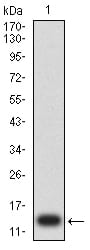
Figure 2:Western blot analysis using CD203C mAb against human CD203C (AA: extra 45-163) recombinant protein. (Expected MW is 13.6 kDa)
Western Blot

Figure 3:Western blot analysis using CD203C mAb against HEK293 (1) and CD203C (AA: extra 45-163)-hIgGFc transfected HEK293 (2) cell lysate.
Flow cytometric

Figure 4:Flow cytometric analysis of HL-60 cells using CD203C mouse mAb (green) and negative control (red).
Immunohistochemical analysis

Figure 5:Immunohistochemical analysis of paraffin-embedded renal cancer tissues using CD203C mouse mAb with DAB staining.
For Research Use Only. Not for use in diagnostic procedures.