

CD2 Primary Antibody
Item Information
Catalog #
Size
Price
Description
CD2 is a surface antigen of the human T-lymphocyte lineage that is expressed on all peripheral blood T cells (summarized by Sewell et al., 1986 [PubMed 3490670]). It is one of the earliest T-cell markers, being present on more than 95% of thymocytes; it is also found on some natural killer cells but not on B lymphocytes. Monoclonal antibodies directed against CD2 inhibit the formation of rosettes with sheep erythrocytes, indicating that CD2 is the erythrocyte receptor or is closely associated with it.
Product Overview
Entrez GenelD
914
Aliases
T11; SRBC; LFA-2
Clone#
3D1E3
Host / Isotype
Mouse / IgG1
Species Reactivity
Mouse
Immunogen
Purified recombinant fragment of human CD2 (AA: 25-140) expressed in E. Coli.
Formulation
Purified antibody in PBS with 0.05% sodium azide
Storage
Store at 4°C short term. Aliquot and store at -20°C long term. Avoid freeze/thaw cycles.
Product Applications
WB (Western Blot)
1/500 - 1/2000
IHC_P(Immunohistochemistry)
1/200 - 1/1000
FCM (Flow Cytometry)
1/200 - 1/400
ELISA
1/10000
References
1.J Virol. 2013 Aug;87(16):9148-58.
2.PLoS One. 2012;7(10):e47664.
2.PLoS One. 2012;7(10):e47664.
Product Image
Elisa

Figure 1: Black line: Control Antigen (100 ng);Purple line: Antigen (10ng); Blue line: Antigen (50 ng); Red line:Antigen (100 ng)
Western Blot

Figure 2:Western blot analysis using CD2 mAb against human CD2 (AA: 25-140) recombinant protein. (Expected MW is 39.2 kDa)
Western Blot

Figure 3:Western blot analysis using CD2 mAb against HEK293 (1) and CD2 (AA: 25-140)-hIgGFc transfected HEK293 (2) cell lysate.
Western Blot

Figure 4:Western blot analysis using CD2 mouse mAb against MOLT4 (1), MCF-7 (2), L1210 (3), U937 (4), and NIH3T3 (5) cell lysate.
Flow cytometric

Figure 5:Flow cytometric analysis of Hela cells using CD2 mouse mAb (green) and negative control (red).
Flow cytometric
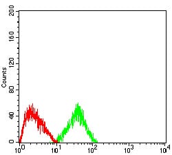
Figure 6:Flow cytometric analysis of HepG2 cells using CD2 mouse mAb (green) and negative control (red).
Immunohistochemical analysis

Figure 7:Immunohistochemical analysis of paraffin-embedded rectum cancer tissues using CD2 mouse mAb with DAB staining.
Immunohistochemical analysis

Figure 8:Immunohistochemical analysis of paraffin-embedded endometrial cancer tissues using CD2 mouse mAb with DAB staining.
For Research Use Only. Not for use in diagnostic procedures.