

CD1A Primary Antibody
Item Information
Catalog #
Size
Price
Description
CD1a is a non polymorphic MHC Class 1 related cell surface glycoprotein, expressed in association with Beta 2 microglobulin. CD1a is expressed by cortical thymocytes, Langerhan's cells and by interdigitating cells. CD1a is also expressed by some malignancies of T cell lineage and in histiocytosis X. Tissue specificity: Expressed on cortical thymocytes, epidermal Langerhans cells, dendritic cells, on certain T-cell leukemias, and in various other tissues.
Product Overview
Entrez GenelD
909
Aliases
R4; T6; CD1; FCB6; HTA1; CD1A
Clone#
7A7
Host / Isotype
Mouse / IgG1
Species Reactivity
Human
Immunogen
Purified recombinant fragment of human CD1A expressed in E. Coli.
Formulation
Ascitic fluid containing 0.03% sodium azide.
Storage
Store at 4°C short term. Aliquot and store at -20°C long term. Avoid freeze/thaw cycles.
Product Applications
WB (Western Blot)
1/500 - 1/2000
IHC_P(Immunohistochemistry)
1/200 - 1/1000
ICC (Immunocytochemistry)
1/200 - 1/1000
ELISA
1/10000
References
1. J Neuroimmunol. 2008 Dec 15;205(1-2):110-2.
2. Pathol Int. 2008 Mar;58(3):169-73.
2. Pathol Int. 2008 Mar;58(3):169-73.
Product Image
Western Blot

Figure 1: Western blot analysis using CD1A mouse mAb against K562 (1), RAJI (2), and MOLT4 (3) cell lysate.
Immunohistochemical analysis
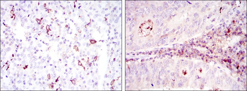
Figure 2: Immunohistochemical analysis of paraffin-embedded cervical cancer tissues (left) and colon cancer tissues (right) using CD1A mouse mAb with DAB staining.
Immunohistochemical analysis

Figure 3: Immunohistochemical analysis of paraffin-embedded brain tissues (left) and submaxillary tumor tissues (right) using CD1A mouse mAb with DAB staining.
Immunofluorescence analysis

Figure 4: Immunofluorescence analysis of RAJI cells using CD1A mouse mAb (green). Blue: DRAQ5 fluorescent DNA dye.
Elisa

Red: Control Antigen (100ng); Purple: Antigen (10ng); Green: Antigen (50ng); Blue: Antigen (100ng);
For Research Use Only. Not for use in diagnostic procedures.