

CD195 Primary Antibody
Item Information
Catalog #
Size
Price
Description
This gene encodes a member of the beta chemokine receptor family, which is predicted to be a seven transmembrane protein similar to G protein-coupled receptors. This protein is expressed by T cells and macrophages, and is known to be an important co-receptor for macrophage-tropic virus, including HIV, to enter host cells. Defective alleles of this gene have been associated with the HIV infection resistance. The ligands of this receptor include monocyte chemoattractant protein 2 (MCP-2), macrophage inflammatory protein 1 alpha (MIP-1 alpha), macrophage inflammatory protein 1 beta (MIP-1 beta) and regulated on activation normal T expressed and secreted protein (RANTES). Expression of this gene was also detected in a promyeloblastic cell line, suggesting that this protein may play a role in granulocyte lineage proliferation and differentiation. This gene is located at the chemokine receptor gene cluster region. An allelic polymorphism in this gene results in both functional and non-functional alleles; the reference genome represents the functional allele. Two transcript variants encoding the same protein have been found for this gene.
Product Overview
Entrez GenelD
1234
Aliases
CCR5; CKR5; CCR-5; CKR-5; CCCKR5; CMKBR5; IDDM22; CC-CKR-5
Clone#
6G11D1
Host / Isotype
Mouse / IgG1
Species Reactivity
Human, Rat
Immunogen
Purified recombinant fragment of human CD195 (AA: extra mix) expressed in E. Coli.
Formulation
Purified antibody in PBS with 0.05% sodium azide
Storage
Store at 4°C short term. Aliquot and store at -20°C long term. Avoid freeze/thaw cycles.
Product Applications
WB (Western Blot)
1/500 - 1/2000
IHC_P(Immunohistochemistry)
1/200 - 1/1000
FCM (Flow Cytometry)
1/200 - 1/400
ELISA
1/10000
References
1.J Leukoc Biol. 2015 Jul;98(1):59-71.
2.J Gen Virol. 2015 Aug;96(8):2074-8.
2.J Gen Virol. 2015 Aug;96(8):2074-8.
Product Image
Elisa
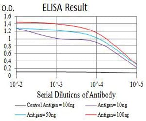
Figure 1: Black line: Control Antigen (100 ng);Purple line: Antigen (10ng); Blue line: Antigen (50 ng); Red line:Antigen (100 ng)
Western Blot

Figure 2:Western blot analysis using CD195 mAb against human CD195 (AA: extra mix) recombinant protein. (Expected MW is 36.7 kDa)
Western Blot

Figure 3:Western blot analysis using CD195 mAb against HEK293 (1) and CD195 (AA: extra mix)-hIgGFc transfected HEK293 (2) cell lysate.
Western Blot

Figure 4:Western blot analysis using CD195 mouse mAb against MOLT4 (1), L-02 (2), SPA-C-1 (3), A549 (4), and C6 (5) cell lysate.
Flow cytometric

Figure 5:Flow cytometric analysis of K562 cells using CD195 mouse mAb (green) and negative control (red).
Immunohistochemical analysis

Figure 6:Immunohistochemical analysis of paraffin-embedded cervical cancer tissues using CD195 mouse mAb with DAB staining.
Immunohistochemical analysis

Figure 7:Immunohistochemical analysis of paraffin-embedded bladder cancer tissues using CD195 mouse mAb with DAB staining.
For Research Use Only. Not for use in diagnostic procedures.