

CD192 Primary Antibody
Item Information
Catalog #
Size
Price
Description
The protein encoded by this gene is a receptor for monocyte chemoattractant protein-1, a chemokine which specifically mediates monocyte chemotaxis. Monocyte chemoattractant protein-1 is involved in monocyte infiltration in inflammatory diseases such as rheumatoid arthritis as well as in the inflammatory response against tumors. The encoded protein mediates agonist-dependent calcium mobilization and inhibition of adenylyl cyclase. This protein can also be a coreceptor with CD4 for HIV-1 infection. This gene is located in the chemokine receptor gene cluster region of chromosome 3.
Product Overview
Entrez GenelD
729230
Aliases
CCR2; CKR2; CCR-2; CCR2A; CCR2B; CKR2A; CKR2B; CMKBR2; MCP-1-R; CC-CKR-2
Clone#
3B6B1
Host / Isotype
Mouse / IgG1
Species Reactivity
Human
Immunogen
Purified recombinant fragment of human CD192 expressed in E. Coli.
Formulation
Purified antibody in PBS with 0.05% sodium azide
Storage
Store at 4°C short term. Aliquot and store at -20°C long term. Avoid freeze/thaw cycles.
Product Applications
WB (Western Blot)
1/500 - 1/2000
FCM (Flow Cytometry)
1/200 - 1/400
ELISA
1/10000
References
1.PLoS One. 2014 Nov 20;9(11):e113304.
2.J Cereb Blood Flow Metab. 2014 Sep;34(9):1425-9.
2.J Cereb Blood Flow Metab. 2014 Sep;34(9):1425-9.
Product Image
Elisa
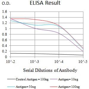
Figure 1:Black line: Control Antigen (100 ng);Purple line: Antigen (10ng); Blue line: Antigen (50 ng); Red line:Antigen (100 ng)
Western Blot
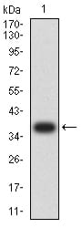
Figure 2:Western blot analysis using CD192 mAb against human CD192 recombinant protein. (Expected MW is 37.6 kDa)
Western Blot
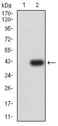
Figure 3:Western blot analysis using CD192 mAb against HEK293 (1) and CD192-hIgGFc transfected HEK293 (2) cell lysate.
Flow cytometric
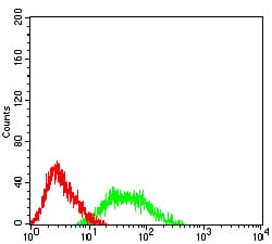
Figure 4:Flow cytometric analysis of HL-60 cells using CD192 mouse mAb (green) and negative control (red).
For Research Use Only. Not for use in diagnostic procedures.