

CD183 Primary Antibody
Item Information
Catalog #
Size
Price
Description
This gene encodes a G protein-coupled receptor with selectivity for three chemokines, termed CXCL9/Mig (monokine induced by interferon-g), CXCL10/IP10 (interferon-g-inducible 10 kDa protein) and CXCL11/I-TAC (interferon-inducible T cell a-chemoattractant). Binding of chemokines to this protein induces cellular responses that are involved in leukocyte traffic, most notably integrin activation, cytoskeletal changes and chemotactic migration. Alternatively spliced transcript variants encoding different isoforms have been found for this gene. One of the isoforms (CXCR3-B) shows high affinity binding to chemokine, CXCL4/PF4 (PMID:12782716).
Product Overview
Entrez GenelD
2833
Aliases
CXCR3; GPR9; MigR; CD182; Mig-R; CKR-L2; CMKAR3; IP10-R
Clone#
5C10B3
Host / Isotype
Mouse / IgG1
Species Reactivity
Human
Immunogen
Purified recombinant fragment of human CD183 (AA: extra mix) expressed in E. Coli.
Formulation
Purified antibody in PBS with 0.05% sodium azide
Storage
Store at 4°C short term. Aliquot and store at -20°C long term. Avoid freeze/thaw cycles.
Product Applications
WB (Western Blot)
1/500 - 1/2000
IHC_P(Immunohistochemistry)
1/200 - 1/1000
FCM (Flow Cytometry)
1/200 - 1/400
ELISA
1/10000
References
1.Hum Pathol. 2015 Dec;46(12):1872-80.
2.Breast Cancer Res Treat. 2015 Jan;149(2):403-15.
2.Breast Cancer Res Treat. 2015 Jan;149(2):403-15.
Product Image
Elisa

Figure 1: Black line: Control Antigen (100 ng);Purple line: Antigen (10ng); Blue line: Antigen (50 ng); Red line:Antigen (100 ng)
Western Blot

Figure 2:Western blot analysis using CD183 mAb against human CD183 (AA: extra mix) recombinant protein. (Expected MW is 38.3 kDa)
Western Blot

Figure 3:Western blot analysis using CD183 mAb against HEK293 (1) and CD183 (AA: extra mix)-hIgGFc transfected HEK293 (2) cell lysate.
Western Blot

Figure 4:Western blot analysis using CD183 mouse mAb against Hela (1) and L-02 (2) cell lysate.
Flow cytometric
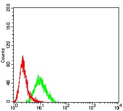
Figure 5:Flow cytometric analysis of HL-60 cells using CD183 mouse mAb (green) and negative control (red).
Flow cytometric
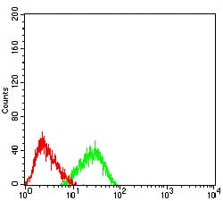
Figure 6:Flow cytometric analysis of Jurkat cells using CD183 mouse mAb (green) and negative control (red).
Immunohistochemical analysis

Figure 7:Immunohistochemical analysis of paraffin-embedded bladder cancer tissues using CD183 mouse mAb with DAB staining.
For Research Use Only. Not for use in diagnostic procedures.