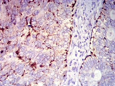
CD182 Primary Antibody

Item Information
Catalog #
Size
Price
Description

The protein encoded by this gene is a member of the G-protein-coupled receptor family. This protein is a receptor for interleukin 8 (IL8). It binds to IL8 with high affinity, and transduces the signal through a G-protein activated second messenger system. This receptor also binds to chemokine (C-X-C motif) ligand 1 (CXCL1/MGSA), a protein with melanoma growth stimulating activity, and has been shown to be a major component required for serum-dependent melanoma cell growth. This receptor mediates neutrophil migration to sites of inflammation. The angiogenic effects of IL8 in intestinal microvascular endothelial cells are found to be mediated by this receptor. Knockout studies in mice suggested that this receptor controls the positioning of oligodendrocyte precursors in developing spinal cord by arresting their migration. This gene, IL8RA, a gene encoding another high affinity IL8 receptor, as well as IL8RBP, a pseudogene of IL8RB, form a gene cluster in a region mapped to chromosome 2q33-q36. Alternatively spliced variants, encoding the same protein, have been identified.

Product Overview
Entrez GenelD
3579
Aliases
CXCR2; IL8R2; IL8RA; IL8RB; CMKAR2; CDw128b
Clone#
7E9H12
Host / Isotype
Mouse / IgG1
Species Reactivity
Human
Immunogen
Purified recombinant fragment of human CD182 expressed in E. Coli.
Formulation
Purified antibody in PBS with 0.05% sodium azide
Storage
4°C; -20°C for long term storage
Product Applications
WB (Western Blot)
1/500 - 1/2000
IHC_P(Immunohistochemistry)
1/200 - 1/1000
FCM (Flow Cytometry)
1/200 - 1/400
ELISA
1/10000
References
1.Oncotarget. 2017 Apr 25;8(17):28442-28454.
2.Eur J Cancer. 2015 Sep;51(14):1953-61.
Product Image
Elisa
Figure 1:Black line: Control Antigen (100 ng);Purple line: Antigen (10ng); Blue line: Antigen (50 ng); Red line:Antigen (100 ng)
Western Blot
Figure 2:Western blot analysis using CD182 mAb against human CD182 recombinant protein. (Expected MW is 38.6 kDa)
Western Blot
Figure 3:Western blot analysis using CD182 mAb against HEK293 (1) and CD182-hIgGFc transfected HEK293 (2) cell lysate.
Flow cytometric
Figure 4:Flow cytometric analysis of Ramos cells using CD182 mouse mAb (green) and negative control (red).
Immunohistochemical analysis
Figure 5:Immunohistochemical analysis of paraffin-embedded cervical cancer tissues using CD182 mouse mAb with DAB staining.
For Research Use Only. Not for use in diagnostic procedures.