
CD18 Primary Antibody
Item Information
Catalog #
Size
Price
Description
This gene encodes an integrin beta chain, which combines with multiple different alpha chains to form different integrin heterodimers. Integrins are integral cell-surface proteins that participate in cell adhesion as well as cell-surface mediated signalling. The encoded protein plays an important role in immune response and defects in this gene cause leukocyte adhesion deficiency. Alternative splicing results in multiple transcript variants.
Product Overview
Entrez GenelD
3689
Aliases
ITGB2; LAD; CD18; MF17; MFI7; LCAMB; LFA-1; MAC-1
Clone#
3D1B3
Host / Isotype
Mouse / IgG1
Species Reactivity
Human
Immunogen
Purified recombinant fragment of human CD18 (AA: extra 559-700) expressed in E. Coli.
Formulation
Purified antibody in PBS with 0.05% sodium azide
Storage
Store at 4°C short term. Aliquot and store at -20°C long term. Avoid freeze/thaw cycles.
Product Applications
WB (Western Blot)
1/500 - 1/2000
FCM (Flow Cytometry)
1/200 - 1/400
ELISA
1/10000
References
1.Blood. 2017 Jul 6;130(1):86-88.
2.Ann Hematol. 2016 Dec;95(12):1965-1969.
2.Ann Hematol. 2016 Dec;95(12):1965-1969.
Product Image
Elisa

Figure 1:Black line: Control Antigen (100 ng);Purple line: Antigen (10ng); Blue line: Antigen (50 ng); Red line:Antigen (100 ng)
Western Blot

Figure 2:Western blot analysis using CD18 mAb against human CD18 (AA: extra 559-700) recombinant protein. (Expected MW is 41.5 kDa)
Western Blot

Figure 3:Western blot analysis using CD18 mAb against HEK293 (1) and CD18 (AA: extra 559-700)-hIgGFc transfected HEK293 (2) cell lysate.
Western Blot

Figure 4:Western blot analysis using CD18 mouse mAb against HL-60 (1), Jurkat (2), and MOLT4 (3) cell lysate.
Flow cytometric
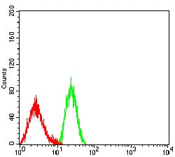
Figure 5:Flow cytometric analysis of HL-60 cells using CD18 mouse mAb (green) and negative control (red).
For Research Use Only. Not for use in diagnostic procedures.