

CD163 Primary Antibody
Item Information
Catalog #
Size
Price
Description
The protein encoded by this gene is a member of the scavenger receptor cysteine-rich (SRCR) superfamily, and is exclusively expressed in monocytes and macrophages. It functions as an acute phase-regulated receptor involved in the clearance and endocytosis of hemoglobin/haptoglobin complexes by macrophages, and may thereby protect tissues from free hemoglobin-mediated oxidative damage. This protein may also function as an innate immune sensor for bacteria and inducer of local inflammation. Alternatively spliced transcript variants encoding different isoforms have been described for this gene.
Product Overview
Entrez GenelD
9332
Aliases
M130; MM130; SCARI1
Clone#
7G5E2
Host / Isotype
Mouse / Mouse IgG1
Immunogen
Purified recombinant fragment of human CD163 (AA: extra 42-259) expressed in E. Coli.
Formulation
Purified antibody in PBS with 0.05% sodium azide
Storage
Store at 4°C short term. Aliquot and store at -20°C long term. Avoid freeze/thaw cycles.
Product Applications
WB (Western Blot)
1/500 - 1/2000
IHC_P(Immunohistochemistry)
1/200 - 1/1000
FCM (Flow Cytometry)
1/200 - 1/400
ELISA
1/10000
References
1.J Cancer Res Clin Oncol. 2018 Jul;144(7):1253-1263. 2.Cancer Biomark. 2018 Feb 14;21(3):689-700.
Product Image
Elisa

Figure 1:Black line: Control Antigen (100 ng);Purple line: Antigen (10ng); Blue line: Antigen (50 ng); Red line:Antigen (100 ng)
Western Blot

Figure 2:Western blot analysis using CD163 mAb against human CD163 (AA: extra 42-259) recombinant protein. (Expected MW is 26.4 kDa)
Western Blot
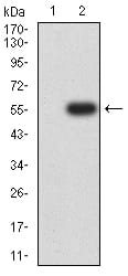
Figure 3:Western blot analysis using CD163 mAb against HEK293 (1) and CD163 (AA: extra 42-259)-hIgGFc transfected HEK293 (2) cell lysate.
Western Blot

Figure 4:Western blot analysis using CD163 mouse mAb against Raw264.7 (1), NIH/3T3 (2), and HL-60 (3) cell lysate.
Immunohistochemical Analysis

Figure 5:Immunohistochemical analysis of paraffin-embedded rectum cancer tissues using CD163 mouse mAb with DAB staining.
For Research Use Only. Not for use in diagnostic procedures.