

CD161 Primary Antibody
Item Information
Catalog #
Size
Price
Description
Natural killer (NK) cells are lymphocytes that mediate cytotoxicity and secrete cytokines after immune stimulation. Several genes of the C-type lectin superfamily, including the rodent NKRP1 family of glycoproteins, are expressed by NK cells and may be involved in the regulation of NK cell function. The KLRB1 protein contains an extracellular domain with several motifs characteristic of C-type lectins, a transmembrane domain, and a cytoplasmic domain. The KLRB1 protein is classified as a type II membrane protein because it has an external C terminus.
Product Overview
Entrez GenelD
3820
Aliases
KLRB1; NKR; CLEC5B; NKR-P1; NKRP1A; NKR-P1A; hNKR-P1A
Clone#
4C6A11
Host / Isotype
Mouse / IgG2b
Species Reactivity
Human
Immunogen
Purified recombinant fragment of human CD161 (AA: extra 67-225) expressed in E. Coli.
Formulation
Purified antibody in PBS with 0.05% sodium azide
Storage
Store at 4°C short term. Aliquot and store at -20°C long term. Avoid freeze/thaw cycles.
Product Applications
WB (Western Blot)
1/500 - 1/2000
FCM (Flow Cytometry)
1/200 - 1/400
ELISA
1/10000
References
1.Biol Blood Marrow Transplant. 2015 Mar;21(3):421-8.
2.Cell Immunol. 2011;269(2):74-7.
2.Cell Immunol. 2011;269(2):74-7.
Product Image
Elisa
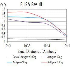
Figure 1: Black line: Control Antigen (100 ng);Purple line: Antigen (10ng); Blue line: Antigen (50 ng); Red line:Antigen (100 ng)
Western Blot

Figure 2:Western blot analysis using CD161 mAb against human CD161 (AA: extra 67-225) recombinant protein. (Expected MW is 48.4 kDa)
Western Blot
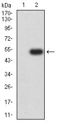
Figure 3:Western blot analysis using CD161 mAb against HEK293 (1) and CD161 (AA: extra 67-225)-hIgGFc transfected HEK293 (2) cell lysate.
Flow cytometric

Figure 4:Flow cytometric analysis of Raji cells using CD161 mouse mAb (green) and negative control (red).
For Research Use Only. Not for use in diagnostic procedures.