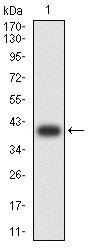
CD160

Item Information
Catalog #
Size
Price
Description

CD160 is an 27 kDa glycoprotein which was initially identified with the monoclonal antibody BY55. Its expression is tightly associated with peripheral blood NK cells and CD8 T lymphocytes with cytolytic effector activity. The cDNA sequence of CD160 predicts a cysteine-rich, glycosylphosphatidylinositol-anchored protein of 181 amino acids with a single Ig-like domain weakly homologous to KIR2DL4 molecule. CD160 is expressed at the cell surface as a tightly disulfide-linked multimer. RNA blot analysis revealed CD160 mRNAs of 1.5 and 1.6 kb whose expression was highly restricted to circulating NK and T cells, spleen and small intestine. Within NK cells CD160 is expressed by CD56dimCD16+ cells whereas among circulating T cells its expression is mainly restricted to TCRgd bearing cells and to TCRab+CD8brightCD95+CD56+CD28-CD27-cells. In tissues, CD160 is expressed on all intestinal intraepithelial lymphocytes. CD160 shows a broad specificity for binding to both classical and nonclassical MHC class I molecules.

Product Overview
Formulation
Purified antibody in PBS with 0.05% sodium azide
Storage
4℃; -20℃ for long term storage
Product Image
Elisa
Figure 1:Black line: Control Antigen (100 ng);Purple line: Antigen (10ng); Blue line: Antigen (50 ng); Red line:Antigen (100 ng)
Western Blot
Figure 2:Western blot analysis using CD160 mAb against human CD160 (AA: 25-155) recombinant protein. (Expected MW is 40.5 kDa)
Western Blot
Figure 3:Western blot analysis using CD160 mAb against HEK293-6e (1) and CD160 (AA: 25-155)-hIgGFc transfected HEK293-6e (2) cell lysate.
Immunofluorescence analysis
Figure 4:Flow cytometric analysis of Jurkat cells using CD160 mouse mAb (green) and negative control (red).
For Research Use Only. Not for use in diagnostic procedures.