
CD16 Primary Antibody
Item Information
Catalog #
Size
Price
Description
This gene encodes a receptor for the Fc portion of immunoglobulin G, and it is involved in the removal of antigen-antibody complexes from the circulation, as well as other other antibody-dependent responses. This gene (FCGR3A) is highly similar to another nearby gene (FCGR3B) located on chromosome 1. The receptor encoded by this gene is expressed on natural killer (NK) cells as an integral membrane glycoprotein anchored through a transmembrane peptide, whereas FCGR3B is expressed on polymorphonuclear neutrophils (PMN) where the receptor is anchored through a phosphatidylinositol (PI) linkage. Mutations in this gene have been linked to susceptibility to recurrent viral infections, susceptibility to systemic lupus erythematosus, and alloimmune neonatal neutropenia. Alternatively spliced transcript variants encoding different isoforms have been found for this gene.
Product Overview
Entrez GenelD
2214
Aliases
FCGR3A; FCG3; CD16A; FCGR3; IGFR3; IMD20; FCR-10; FCRIII; FCGRIII; FCRIIIA
Clone#
2G10A9
Host / Isotype
Mouse / IgG1
Species Reactivity
Human
Immunogen
Purified recombinant fragment of human CD16 (AA: extra 17-208) expressed in E. Coli.
Formulation
Purified antibody in PBS with 0.05% sodium azide
Storage
Store at 4°C short term. Aliquot and store at -20°C long term. Avoid freeze/thaw cycles.
Product Applications
WB (Western Blot)
1/500 - 1/2000
FCM (Flow Cytometry)
1/200 - 1/400
ELISA
1/10000
References
1.Hum Immunol. 2016 Feb;77(2):165-71.
2.PLoS One. 2015 Oct 7;10(10):e0140120.
2.PLoS One. 2015 Oct 7;10(10):e0140120.
Product Image
Elisa
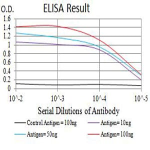
Figure 1: Black line: Control Antigen (100 ng);Purple line: Antigen (10ng); Blue line: Antigen (50 ng); Red line:Antigen (100 ng)
Western Blot

Figure 2:Western blot analysis using CD16 mAb against human CD16 (AA: extra 17-208) recombinant protein. (Expected MW is 47.8 kDa)
Western Blot

Figure 3:Western blot analysis using CD16 mAb against HEK293 (1) and CD16 (AA: extra 17-208)-hIgGFc transfected HEK293 (2) cell lysate.
Flow cytometric

Figure 4:Flow cytometric analysis of Ramos cells using CD16 mouse mAb (green) and negative control (red).
For Research Use Only. Not for use in diagnostic procedures.