

CD131 Primary Antibody
Item Information
Catalog #
Size
Price
Description
The protein encoded by this gene is the common beta chain of the high affinity receptor for IL-3, IL-5 and CSF. Defects in this gene have been reported to be associated with protein alveolar proteinosis (PAP).
Product Overview
Entrez GenelD
1439
Aliases
CSF2RB; IL3RB; IL5RB; SMDP5; CDw131; betaGMR
Clone#
1D5E4
Host / Isotype
Mouse / Mouse IgG1
Immunogen
Purified recombinant fragment of human CD131 (AA: extra 17-149) expressed in E. Coli.
Formulation
Purified antibody in PBS with 0.05% sodium azide
Storage
Store at 4°C short term. Aliquot and store at -20°C long term. Avoid freeze/thaw cycles.
Product Applications
WB (Western Blot)
1/500 - 1/2000
IHC_P(Immunohistochemistry)
1/200 - 1/1000
FCM (Flow Cytometry)
1/200 - 1/400
ELISA
1/10000
References
1.Int J Hematol. 2011 Jan;93(1):83-90. 2.Eur Respir J. 2011 Jan;37(1):201-4.
Product Image
Elisa

Figure 1:Black line: Control Antigen (100 ng);Purple line: Antigen (10ng); Blue line: Antigen (50 ng); Red line:Antigen (100 ng)
Western Blot

Figure 2:Western blot analysis using CD131 mAb against human CD131 (AA: 17-149) recombinant protein. (Expected MW is 41.3 kDa)
Western Blot

Figure 3:Western blot analysis using CD131 mAb against HEK293 (1) and CD131 (AA: 17-149)-hIgGFc transfected HEK293 (2) cell lysate.
Western Blot

Figure 4:Western blot analysis using CD131 mouse mAb against NIH/3T3 (1), Hela (2), C6 (3) and MCF-7 (4) cell lysate.
Flow Cytometric

Figure 5:Flow cytometric analysis of Hela cells using CD131 mouse mAb (green) and negative control (red).
Immunohistochemical Analysis
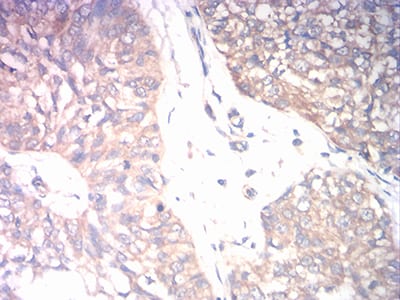
Figure 6:Immunohistochemical analysis of paraffin-embedded bladder cancer tissues using CD131 mouse mAb with DAB staining.
For Research Use Only. Not for use in diagnostic procedures.