CD129 Primary Antibody
Item Information
Catalog #
Size
Price
Description
The protein encoded by this gene is a cytokine receptor that specifically mediates the biological effects of interleukin 9 (IL9). The functional IL9 receptor complex requires this protein as well as the interleukin 2 receptor, gamma (IL2RG), a common gamma subunit shared by the receptors of many different cytokines. The ligand binding of this receptor leads to the activation of various JAK kinases and STAT proteins, which connect to different biologic responses. This gene is located at the pseudoautosomal regions of X and Y chromosomes. Genetic studies suggested an association of this gene with the development of asthma. Multiple pseudogenes on chromosome 9, 10, 16, and 18 have been described. Alternatively spliced transcript variants have been found for this gene.
Product Overview
Entrez GenelD
3581
Aliases
IL9R; IL-9R
Clone#
4A11H2
Host / Isotype
Mouse / IgG1
Species Reactivity
Human, Rat
Immunogen
Purified recombinant fragment of human CD129 (AA: extra 41-270) expressed in E. Coli.
Formulation
Purified antibody in PBS with 0.05% sodium azide
Storage
Store at 4°C short term. Aliquot and store at -20°C long term. Avoid freeze/thaw cycles.
Product Applications
WB (Western Blot)
1/500 - 1/2000
IHC_P(Immunohistochemistry)
1/200 - 1/1000
FCM (Flow Cytometry)
1/200 - 1/400
ELISA
1/10000
References
1.Oncol Rep. 2015 Aug;34(2):795-802.
2.Int J Clin Exp Pathol. 2013 Apr 15;6(5):911-6.
2.Int J Clin Exp Pathol. 2013 Apr 15;6(5):911-6.
Product Image
Elisa

Figure 1: Black line: Control Antigen (100 ng);Purple line: Antigen (10ng); Blue line: Antigen (50 ng); Red line:Antigen (100 ng)
Western Blot

Figure 2:Western blot analysis using CD129 mAb against human CD129 (AA: extra 41-270) recombinant protein. (Expected MW is 51.9 kDa)
Western Blot

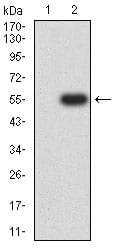
Figure 3:Western blot analysis using CD129 mAb against HEK293 (1) and CD129 (AA: extra 41-270)-hIgGFc transfected HEK293 (2) cell lysate.
Western Blot

Figure 4:Western blot analysis using CD129 mouse mAb against C6 (1) and PC-3 (2) cell lysate.
Flow cytometric

Figure 5:Flow cytometric analysis of Ramos cells using CD129 mouse mAb (green) and negative control (red).
Immunohistochemical analysis

Figure 6:Immunohistochemical analysis of paraffin-embedded cervical cancer tissues using CD129 mouse mAb with DAB staining.
Immunohistochemical analysis

Figure 7:Immunohistochemical analysis of paraffin-embedded ovarian cancer tissues using CD129 mouse mAb with DAB staining.
For Research Use Only. Not for use in diagnostic procedures.

