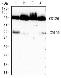
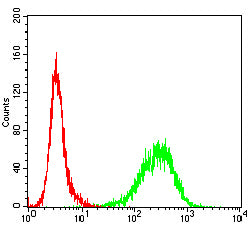
Mouse Monoclonal Antibody to CD126

Item Information
Catalog #
Size
Price
Description

This gene encodes a subunit of the interleukin 6 (IL6) receptor complex. Interleukin 6 is a potent pleiotropic cytokine that regulates cell growth and differentiation and plays an important role in the immune response. The IL6 receptor is a protein complex consisting of this protein and interleukin 6 signal transducer (IL6ST/GP130/IL6-beta), a receptor subunit also shared by many other cytokines. Dysregulated production of IL6 and this receptor are implicated in the pathogenesis of many diseases, such as multiple myeloma, autoimmune diseases and prostate cancer. Alternatively spliced transcript variants encoding distinct isoforms have been identified in this gene. A pseudogene of this gene is found on chromosome 9. [provided by RefSeq, Aug 2020]

Product Overview
Entrez GenelD
3570
Aliases
IL6Q; gp80; CD126; HIES5; IL-6R; IL6RA; IL6RQ; IL-1Ra; IL-6RA; IL6QTL; IL-6R-1
Clone#
4A7B2
Host / Isotype
Mouse / IgG1
Immunogen
Purified recombinant fragment of human CD126 (AA:EXTRA 20-177) expressed in E. Coli.
Formulation
Purified antibody in PBS with 0.05% sodium azide
Storage
4℃; -20℃ for long term storage
Product Applications
WB (Western Blot)
1/500 - 1/2000
FCM (Flow Cytometry)
1/200 - 1/400
ELISA
1/10000
References
1.Medicine (Baltimore).2020 Dec 11;99(50):e23659.2.Biomed Res Int.2021 Jan 28;2021:2615059.
Product Image
Elisa
Figure 1:Black line: Control Antigen (100 ng);Purple line: Antigen (10ng); Blue line: Antigen (50 ng); Red line:Antigen (100 ng)
Western Blot
Figure 2:Western blot analysis using CD126 mAb against human CD126 (AA:20-177) recombinant protein. (Expected MW is 20.3 kDa)
Western Blot
Figure 3:Western blot analysis using CD126 mAb against HEK293 (1) and CD126 (AA:20-177)-hIgGFc transfected HEK293-6e (2) cell lysate.
Western Blot
Figure 4:Western blot analysis using CD126 mouse mAb against Jurkat (1), MOLT4 (2),Raw264.7 (3) and THP-1 (4) cell lysate.
Flow cytometric analysis
Figure 5:Flow cytometric analysis of Jurkat cells using CD126 mouse mAb (green) and negative control (red).
Flow cytometric analysis
Figure 6:Flow cytometric analysis of K562 cells using CD126 mouse mAb (green) and negative control (red).
Flow cytometric analysis
Figure 7:Flow cytometric analysis of THP-1 cells using CD126 mouse mAb (green) and negative control (red).
Flow cytometric analysis
Figure 8:Flow cytometric analysis of U937 cells using CD126 mouse mAb (green) and negative control (red).
For Research Use Only. Not for use in diagnostic procedures.