CD122 Primary Antibody
Item Information
Catalog #
Size
Price
Description
The interleukin 2 receptor, which is involved in T cell-mediated immune responses, is present in 3 forms with respect to ability to bind interleukin 2. The low affinity form is a monomer of the alpha subunit and is not involved in signal transduction. The intermediate affinity form consists of an alpha/beta subunit heterodimer, while the high affinity form consists of an alpha/beta/gamma subunit heterotrimer. Both the intermediate and high affinity forms of the receptor are involved in receptor-mediated endocytosis and transduction of mitogenic signals from interleukin 2. The protein encoded by this gene represents the beta subunit and is a type I membrane protein. The use of alternative promoters results in multiple transcript variants encoding the same protein. The protein is primarily expressed in the hematopoietic system. The use by some variants of an alternate promoter in an upstream long terminal repeat (LTR) results in placenta-specific expression.
Product Overview
Entrez GenelD
3560
Aliases
IL2RB; IL15RB; P70-75
Clone#
1G11C6
Host / Isotype
Mouse / IgG2b
Species Reactivity
Human
Immunogen
Purified recombinant fragment of human CD122 (AA: extra 27-240) expressed in E. Coli.
Formulation
Purified antibody in PBS with 0.05% sodium azide
Storage
Store at 4°C short term. Aliquot and store at -20°C long term. Avoid freeze/thaw cycles.
Product Applications
WB (Western Blot)
1/500 - 1/2000
FCM (Flow Cytometry)
1/200 - 1/400
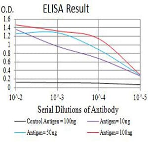
ELISA
1/10000
References
1.Dis Markers. 2014;2014:249846.
2.Am J Physiol Renal Physiol. 2014 May 1;306(9):F1039-46.
2.Am J Physiol Renal Physiol. 2014 May 1;306(9):F1039-46.
Product Image
Elisa

Figure 1: Black line: Control Antigen (100 ng);Purple line: Antigen (10ng); Blue line: Antigen (50 ng); Red line:Antigen (100 ng)
Western Blot

Figure 2:Western blot analysis using CD122 mAb against human CD122 (AA: extra 27-240) recombinant protein. (Expected MW is 50.6 kDa)
Western Blot

Figure 3:Western blot analysis using CD122 mAb against HEK293 (1) and CD122 (AA: extra 27-240)-hIgGFc transfected HEK293 (2) cell lysate.
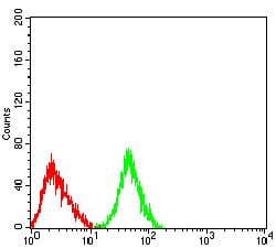
Flow cytometric

Figure 4:Flow cytometric analysis of HL-60 cells using CD122 mouse mAb (green) and negative control (red).
Flow cytometric

Figure 5:Flow cytometric analysis of Jurkat cells using CD122 mouse mAb (green) and negative control (red).
For Research Use Only. Not for use in diagnostic procedures.

