

CD120B Primary Antibody
Item Information
Catalog #
Size
Price
Description
The protein encoded by this gene is a member of the TNF-receptor superfamily. This protein and TNF-receptor 1 form a heterocomplex that mediates the recruitment of two anti-apoptotic proteins, c-IAP1 and c-IAP2, which possess E3 ubiquitin ligase activity. The function of IAPs in TNF-receptor signalling is unknown, however, c-IAP1 is thought to potentiate TNF-induced apoptosis by the ubiquitination and degradation of TNF-receptor-associated factor 2, which mediates anti-apoptotic signals. Knockout studies in mice also suggest a role of this protein in protecting neurons from apoptosis by stimulating antioxidative pathways.
Product Overview
Entrez GenelD
7133
Aliases
TNFRSF1B; p75; TBPII; TNFBR; TNFR2; TNFR1B; TNFR80; TNF-R75; p75TNFR; TNF-R-II
Clone#
2H11C2
Host / Isotype
Mouse / IgG2b
Species Reactivity
Human, Mouse
Immunogen
Purified recombinant fragment of human CD120B (AA: extra 23-257) expressed in E. Coli.
Formulation
Purified antibody in PBS with 0.05% sodium azide
Storage
Store at 4°C short term. Aliquot and store at -20°C long term. Avoid freeze/thaw cycles.
Product Applications
WB (Western Blot)
1/500 - 1/2000
FCM (Flow Cytometry)
1/200 - 1/400
ELISA
1/10000
References
1.Clin Exp Immunol. 2016 Aug;185(2):263-70.
2.Cancer Immunol Immunother. 2015 Nov;64(11):1475-85.
2.Cancer Immunol Immunother. 2015 Nov;64(11):1475-85.
Product Image
Elisa
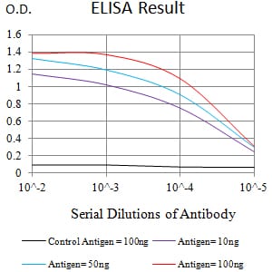
Figure 1:Black line: Control Antigen (100 ng);Purple line: Antigen (10ng); Blue line: Antigen (50 ng); Red line:Antigen (100 ng)
Western Blot
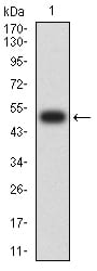
Figure 2:Western blot analysis using CD120B mAb against human CD120B (AA: extra 23-257) recombinant protein. (Expected MW is 51 kDa)
Western Blot
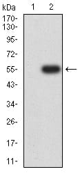
Figure 3:Western blot analysis using CD120B mAb against HEK293 (1) and CD120B (AA: extra 23-257)-hIgGFc transfected HEK293 (2) cell lysate.
Western Blot
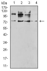
Figure 4:Western blot analysis using CD120B mouse mAb against SK-BR-3 (1), C2C12 (2), MOLT4 (3), and T47D (4) cell lysate.
Flow cytometric
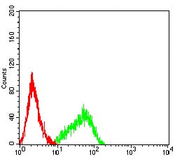
Figure 5:Flow cytometric analysis of HL-60 cells using CD120B mouse mAb (green) and negative control (red).
For Research Use Only. Not for use in diagnostic procedures.