

CD110 Primary Antibody
Item Information
Catalog #
Size
Price
Description
In 1990 an oncogene, v-mpl, was identified from the murine myeloproliferative leukemia virus that was capable of immortalizing bone marrow hematopoietic cells from different lineages. In 1992 the human homologue, named, c-mpl, was cloned. Sequence data revealed that c-mpl encoded a protein that was homologous with members of the hematopoietic receptor superfamily. Presence of anti-sense oligodeoxynucleotides of c-mpl inhibited megakaryocyte colony formation. The ligand for c-mpl, thrombopoietin, was cloned in 1994. Thrombopoietin was shown to be the major regulator of megakaryocytopoiesis and platelet formation. The protein encoded by the c-mpl gene, CD110, is a 635 amino acid transmembrane domain, with two extracellular cytokine receptor domains and two intracellular cytokine receptor box motifs . TPO-R deficient mice were severely thrombocytopenic, emphasizing the important role of CD110 and thrombopoietin in megakaryocyte and platelet formation. Upon binding of thrombopoietin CD110 is dimerized and the JAK family of non-receptor tyrosine kinases, as well as the STAT family, the MAPK family, the adaptor protein Shc and the receptors themselves become tyrosine phosphorylated.
Product Overview
Entrez GenelD
4352
Aliases
MPL; MPLV; TPOR; C-MPL; THCYT2
Clone#
1D6B7
Host / Isotype
Mouse / IgG2a
Species Reactivity
Human
Immunogen
Purified recombinant fragment of human CD110 (AA: extra 26-175) expressed in E. Coli.
Formulation
Purified antibody in PBS with 0.05% sodium azide
Storage
Store at 4°C short term. Aliquot and store at -20°C long term. Avoid freeze/thaw cycles.
Product Applications
WB (Western Blot)
1/500 - 1/2000
FCM (Flow Cytometry)
1/200 - 1/400
ELISA
1/10000
References
1.Haematologica. 2015 Sep;100(9):e341-4.
2.Blood. 2012 Jul 26;120(4):868-79.
2.Blood. 2012 Jul 26;120(4):868-79.
Product Image
Elisa
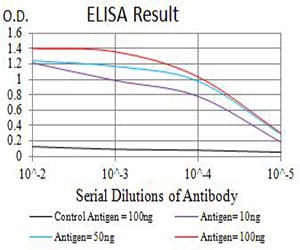
Figure 1: Black line: Control Antigen (100 ng);Purple line: Antigen (10ng); Blue line: Antigen (50 ng); Red line:Antigen (100 ng)
Western Blot
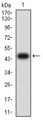
Figure 2:Western blot analysis using CD110 mAb against human CD110 (AA: extra 26-175) recombinant protein. (Expected MW is 47.2 kDa)
Western Blot
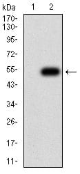
Figure 3:Western blot analysis using CD110 mAb against HEK293 (1) and CD110 (AA: extra 26-175)-hIgGFc transfected HEK293 (2) cell lysate.
Flow cytometric
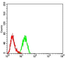
Figure 4:Flow cytometric analysis of K562 cells using CD110 mouse mAb (green) and negative control (red).
For Research Use Only. Not for use in diagnostic procedures.