

CD104 Primary Antibody
Item Information
Catalog #
Size
Price
Description
Integrins are heterodimers comprised of alpha and beta subunits, that are noncovalently associated transmembrane glycoprotein receptors. Different combinations of alpha and beta polypeptides form complexes that vary in their ligand-binding specificities. Integrins mediate cell-matrix or cell-cell adhesion, and transduced signals that regulate gene expression and cell growth. This gene encodes the integrin beta 4 subunit, a receptor for the laminins. This subunit tends to associate with alpha 6 subunit and is likely to play a pivotal role in the biology of invasive carcinoma. Mutations in this gene are associated with epidermolysis bullosa with pyloric atresia. Multiple alternatively spliced transcript variants encoding distinct isoforms have been found for this gene.
Product Overview
Entrez GenelD
3691
Aliases
ITGB4; GP150
Clone#
1D6B4
Host / Isotype
Mouse / IgG2a
Species Reactivity
Human
Immunogen
Purified recombinant fragment of human CD104 (AA: extra 29-206) expressed in E. Coli.
Formulation
Purified antibody in PBS with 0.05% sodium azide
Storage
Store at 4°C short term. Aliquot and store at -20°C long term. Avoid freeze/thaw cycles.
Product Applications
WB (Western Blot)
1/500 - 1/2000
IHC_P(Immunohistochemistry)
1/200 - 1/1000
ICC (Immunocytochemistry)
1/200 - 1/1000
FCM (Flow Cytometry)
1/200 - 1/400
ELISA
1/10000
References
1.Sci Rep. 2015 Nov 17;5:16529.
2.Acta Derm Venereol. 2015 Jan;95(1):112-3.
2.Acta Derm Venereol. 2015 Jan;95(1):112-3.
Product Image
Elisa

Figure 1: Black line: Control Antigen (100 ng);Purple line: Antigen (10ng); Blue line: Antigen (50 ng); Red line:Antigen (100 ng)
Western Blot

Figure 2:Western blot analysis using CD104 mAb against human CD104 (AA: extra 29-206) recombinant protein. (Expected MW is 46.5 kDa)
Western Blot

Figure 3:Western blot analysis using CD104 mAb against HEK293 (1) and CD104 (AA: extra 29-206)-hIgGFc transfected HEK293 (2) cell lysate.
Flow cytometric

Figure 4:Flow cytometric analysis of HL-60 cells using CD104 mouse mAb (green) and negative control (red).
Flow cytometric

Figure 5:Flow cytometric analysis of K562 cells using CD104 mouse mAb (green) and negative control (red).
Immunohistochemical analysis
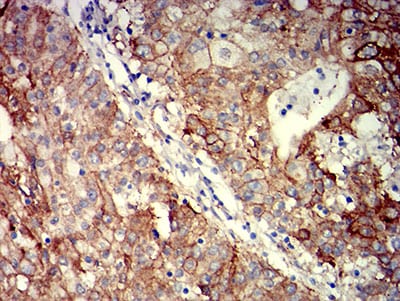
Figure 6:Immunohistochemical analysis of paraffin-embedded stomach cancer tissues using CD104 mouse mAb with DAB staining.
For Research Use Only. Not for use in diagnostic procedures.