

CD10 Primary Antibody
Item Information
Catalog #
Size
Price
Description
The protein encoded by this gene is a type II transmembrane glycoprotein and a common acute lymphocytic leukemia antigen that is an important cell surface marker in the diagnosis of human acute lymphocytic leukemia (ALL). The encoded protein is present on leukemic cells of pre-B phenotype, which represent 85% of cases of ALL. This protein is not restricted to leukemic cells, however, and is found on a variety of normal tissues. The protein is a neutral endopeptidase that cleaves peptides at the amino side of hydrophobic residues and inactivates several peptide hormones including glucagon, enkephalins, substance P, neurotensin, oxytocin, and bradykinin.
Product Overview
Entrez GenelD
4311
Aliases
MME; NEP; SFE; CALLA; CMT2T; SCA43
Clone#
2D12B3
Host / Isotype
Mouse / Mouse IgG1
Immunogen
Purified recombinant fragment of human CD10 (AA: extra 549-750) expressed in E. Coli.
Formulation
Purified antibody in PBS with 0.05% sodium azide
Storage
Store at 4°C short term. Aliquot and store at -20°C long term. Avoid freeze/thaw cycles.
Product Applications
WB (Western Blot)
1/500 - 1/2000
FCM (Flow Cytometry)
1/200-1/400
ELISA
1/10000
References
1.Anticancer Res. 2019 Feb;39(2):635-640. 2.Exp Mol Pathol. 2018 Jun;104(3):190-198.
Product Image
ELISA

Figure 1: Black line: Control Antigen (100 ng);Purple line: Antigen (10ng); Blue line: Antigen (50 ng); Red line: Antigen (100 ng)
WESTERN BLOT

Figure 2: Western blot analysis using CD10 mAb against human CD10 (AA: extra 549-750) recombinant protein. (Expected MW is 48.8 kDa)
WESTERN BLOT

Figure 3: Western blot analysis using CD10 mAb against HEK293-6e (1) and CD10 (AA: extra 549-750)-hIgGFc transfected HEK293-6e (2) cell lysate.
FLOW CYTOMETRY
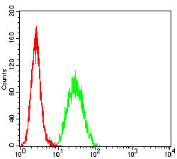
Figure 4: Flow cytometric analysis of Hela cells using CD10 mouse mAb (green) and negative control (red).
For Research Use Only. Not for use in diagnostic procedures.