
CD10 Primary Antibody
Item Information
Catalog #
Size
Price
Description
This gene encodes a common acute lymphocytic leukemia antigen that is an important cell surface marker in the diagnosis of human acute lymphocytic leukemia (ALL). This protein is present on leukemic cells of pre-B phenotype, which represent 85% of cases of ALL. This protein is not restricted to leukemic cells, however, and is found on a variety of normal tissues. It is a glycoprotein that is particularly abundant in kidney, where it is present on the brush border of proximal tubules and on glomerular epithelium. The protein is a neutral endopeptidase that cleaves peptides at the amino side of hydrophobic residues and inactivates several peptide hormones including glucagon, enkephalins, substance P, neurotensin, oxytocin, and bradykinin. This gene, which encodes a 100-kD type II transmembrane glycoprotein, exists in a single copy of greater than 45 kb. The 5' untranslated region of this gene is alternatively spliced, resulting in four separate mRNA transcripts. The coding region is not affected by alternative splicing.
Product Overview
Entrez GenelD
4311
Aliases
NEP; SFE; MME; CALLA; CMT2T; SCA43
Clone#
3G6B11
Host / Isotype
Mouse / IgG1
Species Reactivity
Human
Immunogen
Purified recombinant fragment of human CD10 (AA: extra 549-750) expressed in E. Coli.
Formulation
Purified antibody in PBS with 0.05% sodium azide
Storage
Store at 4°C short term. Aliquot and store at -20°C long term. Avoid freeze/thaw cycles.
Product Applications
WB (Western Blot)
1/500 - 1/2000
IHC_P(Immunohistochemistry)
1/200 - 1/1000
FCM (Flow Cytometry)
1/200 - 1/400
ELISA
1/10000
References
1.Pathobiology. 2015;82(6):259-63.
2.Asian Pac J Cancer Prev. 2015;16(8):3147-52.
2.Asian Pac J Cancer Prev. 2015;16(8):3147-52.
Product Image
Elisa
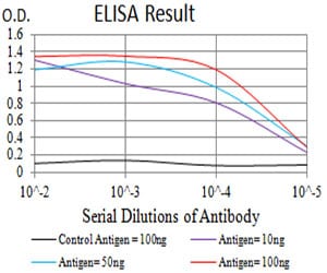
Figure 1: Black line: Control Antigen (100 ng);Purple line: Antigen (10ng); Blue line: Antigen (50 ng); Red line:Antigen (100 ng)
Western Blot

Figure 2:Western blot analysis using CD10 mAb against human CD10 (AA: extra 549-750) recombinant protein. (Expected MW is 48.8 kDa)
Western Blot
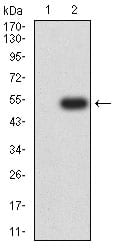
Figure 3:Western blot analysis using CD10 mAb against HEK293 (1) and CD10 (AA: extra 549-750)-hIgGFc transfected HEK293 (2) cell lysate.
Flow cytometric
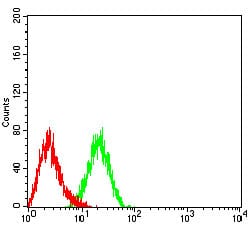
Figure 4:Flow cytometric analysis of HL-60 cells using CD10 mouse mAb (green) and negative control (red).
Immunohistochemical analysis

Figure 5:Immunohistochemical analysis of paraffin-embedded bladder cancer tissues using CD10 mouse mAb with DAB staining.
For Research Use Only. Not for use in diagnostic procedures.