

CCT2 Primary Antibody
Item Information
Catalog #
Size
Price
Description
The protein encoded by this gene is a molecular chaperone that is a member of the chaperonin containing TCP1 complex (CCT), also known as the TCP1 ring complex (TRiC). This complex consists of two identical stacked rings, each containing eight different proteins. Unfolded polypeptides enter the central cavity of the complex and are folded in an ATP-dependent manner. The complex folds various proteins, including actin and tubulin. Two transcript variants encoding different isoforms have been found for this gene.
Product Overview
Entrez GenelD
10576
Aliases
CCTB; 99D8.1; PRO1633; CCT-beta; MGC142074; MGC142076; TCP-1-beta
Clone#
5B5C4
Host / Isotype
Mouse / IgG1
Species Reactivity
Human, Mouse, Monkey, Rat
Immunogen
Purified recombinant fragment of human CCT2 expressed in E. Coli.
Formulation
Ascitic fluid containing 0.03% sodium azide.
Storage
Store at 4°C short term. Aliquot and store at -20°C long term. Avoid freeze/thaw cycles.
Product Applications
WB (Western Blot)
1/500 - 1/2000
ICC (Immunocytochemistry)
1/200 - 1/1000
FCM (Flow Cytometry)
1/200 - 1/400
ELISA
1/10000
References
1. J Biol Chem. 2009 May 29;284(22):14939-48.
2. Mol Cell Proteomics. 2009 Jan;8(1):157-71.
2. Mol Cell Proteomics. 2009 Jan;8(1):157-71.
Product Image
Western Blot

Figure 1: Western blot analysis using CCT2 mAb against human CCT2 (AA: 87-290) recombinant protein. (Expected MW is 47.9 kDa)
Western Blot

Figure 2: Western blot analysis using CCT2 mouse mAb against Hela (1), MCF-7 (2), Jurkat (3), T47D (4), K562 (5), A431 (6), NIH/3T3 (7), PC-12 (8) and Cos7 (9) cell lysate.
Immunofluorescence analysis
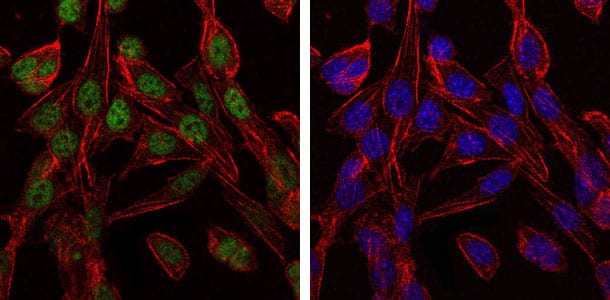
Figure 3: Immunofluorescence analysis of 3T3-L1 cells using CCT2 mouse mAb (green). Blue: DRAQ5 fluorescent DNA dye. Red: Actin filaments have been labeled with Alexa Fluor-555 phalloidin.
Flow cytometric

Figure 4: Flow cytometric analysis of NIH/3T3 cells using CCT2 mouse mAb (blue) and negative control (red).
Elisa

Red: Control Antigen (100ng); Purple: Antigen (10ng); Green: Antigen (50ng); Blue: Antigen (100ng);
For Research Use Only. Not for use in diagnostic procedures.