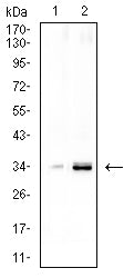
CCND1 Primary Antibody

Item Information
Catalog #
Size
Price
Description

The protein encoded by this gene belongs to the highly conserved cyclin family, whose members are characterized by a dramatic periodicity in protein abundance throughout the cell cycle. Cyclins function as regulators of CDK kinases. Different cyclins exhibit distinct expression and degradation patterns which contribute to the temporal coordination of each mitotic event. This cyclin forms a complex with and functions as a regulatory subunit of CDK4 or CDK6, whose activity is required for cell cycle G1/S transition. This protein has been shown to interact with tumor suppressor protein Rb and the expression of this gene is regulated positively by Rb. Mutations, amplification and overexpression of this gene, which alters cell cycle progression, are observed frequently in a variety of human cancers.

Product Overview
Entrez GenelD
595
Aliases
BCL1; PRAD1; U21B31; D11S287E
Clone#
2A5C9
Host / Isotype
Mouse / Mouse IgG1
Immunogen
Purified recombinant fragment of human CCND1 (AA: 1-295) expressed in E. Coli.
Formulation
Purified antibody in PBS with 0.05% sodium azide
Storage
4°C; -20°C for long term storage
Product Applications
WB (Western Blot)
1/500 - 1/2000
FCM (Flow Cytometry)
1/200-1/400
ELISA
1/10000
References
1.Cancer Med. 2019 Aug;8(9):4100-4109. 2.Sci Rep. 2018 May 1;8(1):6824.
Product Image
ELISA
Figure 1: Black line: Control Antigen (100 ng);Purple line: Antigen (10ng); Blue line: Antigen (50 ng); Red line: Antigen (100 ng)
WESTERN BLOT
Figure 2: Western blot analysis using CCND1 mAb against human CCND1 (AA: 1-295) recombinant protein. (Expected MW is 36.6 kDa)
WESTERN BLOT
Figure 3: Western blot analysis using CCND1 mAb against HEK293-6e (1) and CCND1 (AA: 1-295)-hIgGFc transfected HEK293 (2) cell lysate.
FLOW CYTOMETRY
Figure 4: Flow cytometric analysis of Hela cells using CCND1 mouse mAb (green) and negative control (red).
WESTERN BLOT
Figure 5: Western blot analysis using CCND1 mouse mAb against LNCAP (1), and NIH/3T3 (2) cell lysate.
For Research Use Only. Not for use in diagnostic procedures.