
CCNA2 Primary Antibody
Item Information
Catalog #
Size
Price
Description
The protein encoded by this gene belongs to the highly conserved cyclin family, whose members are characterized by a dramatic periodicity in protein abundance through the cell cycle. Cyclins function as regulators of CDK kinases. Different cyclins exhibit distinct expression and degradation patterns which contribute to the temporal coordination of each mitotic event. In contrast to cyclin A1, which is present only in germ cells, this cyclin is expressed in all tissues tested. This cyclin binds and activates CDC2 or CDK2 kinases, and thus promotes both cell cycle G1/S and G2/M transitions.
Product Overview
Entrez GenelD
890
Aliases
CCN1; CCNA
Clone#
6B4D11
Host / Isotype
Mouse / IgG1
Species Reactivity
Human
Immunogen
Purified recombinant fragment of human CCNA2 (AA: 105-233) expressed in E. Coli.
Formulation
Purified antibody in PBS with 0.05% sodium azide.
Storage
Store at 4°C short term. Aliquot and store at -20°C long term. Avoid freeze/thaw cycles.
Product Applications
WB (Western Blot)
1/500 - 1/2000
IHC_P(Immunohistochemistry)
1/200 - 1/1000
FCM (Flow Cytometry)
1/200 - 1/400
ELISA
1/10000
References
1. Cancer. 2011 Sep 1;117(17):4080-91.
2. J Phys Chem B. 2008 Jul 17;112(28):8346-53.
2. J Phys Chem B. 2008 Jul 17;112(28):8346-53.
Product Image
Western Blot

Figure 1: Western blot analysis using CCNA2 mAb against human CCNA2 (AA: 105-233) recombinant protein. (Expected MW is 40.8 kDa)
Western Blot

Figure 2: Western blot analysis using CCNA2 mAb against HEK293 (1) and CCNA2 (AA: 105-233)-hIgGFc transfected HEK293 (2) cell lysate.
Western Blot

Figure 3: Western blot analysis using CCNA2 mouse mAb against Hela (1), HEK293 (2), Jurkat (3), K562 (4), SK-Br-3 (5), NIH/3T3 (6) cell lysate.
Flow cytometric

Figure 4: Flow cytometric analysis of A431 cells using CCNA2 mouse mAb (green) and negative control (red).
Immunohistochemical analysis

Figure 5: Immunohistochemical analysis of paraffin-embedded cervical cancer tissues using CCNA2 mouse mAb with DAB staining.
Immunohistochemical analysis

Figure 6: Immunohistochemical analysis of paraffin-embedded bladder cancer tissues using CCNA2 mouse mAb with DAB staining.
Elisa
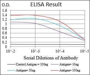
Black line: Control Antigen (100 ng); Purple line: Antigen(10ng); Blue line: Antigen (50 ng); Red line: Antigen (100 ng);
For Research Use Only. Not for use in diagnostic procedures.