

CBX5 Primary Antibody
Item Information
Catalog #
Size
Price
Description
This gene encodes a highly conserved nonhistone protein, which is a member of the heterochromatin protein family. The protein is enriched in the heterochromatin and associated with centromeres. The protein has a single N-terminal chromodomain which can bind to histone proteins via methylated lysine residues, and a C-terminal chromo shadow-domain (CSD) which is responsible for the homodimerization and interaction with a number of chromatin-associated nonhistone proteins. The encoded product is involved in the formation of functional kinetochore through interaction with essential kinetochore proteins. The gene has a pseudogene located on chromosome 3. Multiple alternatively spliced variants, encoding the same protein, have been identified.
Product Overview
Entrez GenelD
23468
Aliases
HP1; HP1A; HEL25
Clone#
2H4E9
Host / Isotype
Mouse / IgG1
Species Reactivity
Human, Mouse
Immunogen
Purified recombinant fragment of human CBX5 (AA: 1-191) expressed in E. Coli.
Formulation
Purified antibody in PBS with 0.05% sodium azide
Storage
Store at 4°C short term. Aliquot and store at -20°C long term. Avoid freeze/thaw cycles.
Product Applications
WB (Western Blot)
1/500 - 1/2000
IHC_P(Immunohistochemistry)
1/200 - 1/1000
ICC (Immunocytochemistry)
1/200 - 1/1000
ELISA
1/10000
References
1.Mol Carcinog. 2011 Aug;50(8):601-13.
2.J Mol Biol. 2013 Jan 9;425(1):54-70.
2.J Mol Biol. 2013 Jan 9;425(1):54-70.
Product Image
Elisa
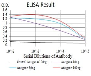
Figure 1: Black line: Control Antigen (100 ng); Purple line: Antigen(10ng); Blue line: Antigen (50 ng); Red line: Antigen (100 ng);
Western Blot
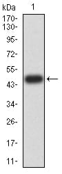
Figure 2:Western blot analysis using CBX5 mAb against human CBX5 (AA: 1-191) recombinant protein. (Expected MW is 48.2 kDa)
Western Blot
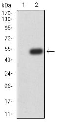
Figure 3:Western blot analysis using CBX5 mAb against HEK293 (1) and CBX5 (AA: 1-191)-hIgGFc transfected HEK293 (2) cell lysate.
Western Blot
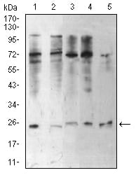
Figure 4:Western blot analysis using CBX5 mouse mAb against Hela (1), NIH/3T3 (2), K562 (3), MCF-7 (4), and A431 (5) cell lysate.
Immunofluorescence analysis
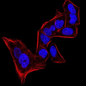
Figure 5:Immunofluorescence analysis of Hela cells using CBX5 mouse mAb. Blue: DRAQ5 fluorescent DNA dye. Red: Actin filaments have been labeled with Alexa Fluor- 555 phalloidin.
Immunofluorescence analysis
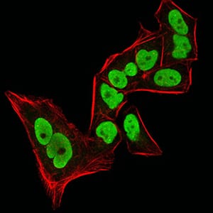
Figure 6:Immunofluorescence analysis of Hela cells using CBX5 mouse mAb (green). Blue: DRAQ5 fluorescent DNA dye. Red: Actin filaments have been labeled with Alexa Fluor- 555 phalloidin. Secondary antibody from Fisher (Cat#: 35503)
Immunohistochemical analysis
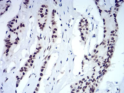
Figure 7:Immunohistochemical analysis of paraffin-embedded colon cancer tissues using CBX5 mouse mAb with DAB staining.
Immunohistochemical analysis
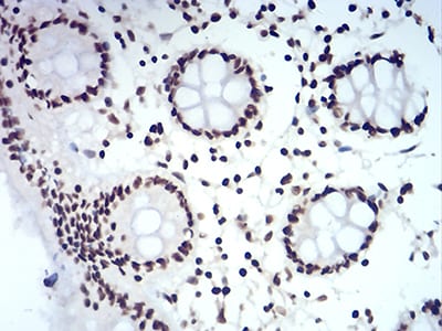
Figure 8:Immunohistochemical analysis of paraffin-embedded colon tissues using CBX5 mouse mAb with DAB staining.
For Research Use Only. Not for use in diagnostic procedures.