

CBX3 Primary Antibody
Item Information
Catalog #
Size
Price
Description
At the nuclear envelope, the nuclear lamina and heterochromatin are adjacent to the inner nuclear membrane. The protein encoded by this gene binds DNA and is a component of heterochromatin. This protein also can bind lamin B receptor, an integral membrane protein found in the inner nuclear membrane. The dual binding functions of the encoded protein may explain the association of heterochromatin with the inner nuclear membrane. This protein binds histone H3 tails methylated at Lys-9 sites. This protein is also recruited to sites of ultraviolet-induced DNA damage and double-strand breaks. Two transcript variants encoding the same protein but differing in the 5' UTR, have been found for this gene.
Product Overview
Entrez GenelD
11335
Aliases
HECH; HP1-GAMMA; HP1Hs-gamma
Clone#
6D1C4
Host / Isotype
Mouse / IgG1
Species Reactivity
Human
Immunogen
Purified recombinant fragment of human CBX3 (AA: 1-183) expressed in E. Coli.
Formulation
Purified antibody in PBS with 0.05% sodium azide
Storage
Store at 4°C short term. Aliquot and store at -20°C long term. Avoid freeze/thaw cycles.
Product Applications
WB (Western Blot)
1/500 - 1/2000
IHC_P(Immunohistochemistry)
1/200 - 1/1000
FCM (Flow Cytometry)
1/200 - 1/400
ELISA
1/10000
References
1.BMC Cancer. 2013 Mar 23;13:148.
2.PLoS One. 2012;7(8):e41401.
2.PLoS One. 2012;7(8):e41401.
Product Image
Elisa

Figure 1: Black line: Control Antigen (100 ng); Purple line: Antigen(10ng); Blue line: Antigen (50 ng); Red line: Antigen (100 ng);
Western Blot

Figure 2:Western blot analysis using CBX3 mAb against human CBX3 (AA: 1-183) recombinant protein. (Expected MW is 46.8 kDa)
Western Blot

Figure 3:Western blot analysis using CBX3 mAb against HEK293 (1) and CBX3 (AA: 1-183)-hIgGFc transfected HEK293 (2) cell lysate.
Flow cytometric

Figure 4:Flow cytometric analysis of A431 cells using CBX3 mouse mAb (green) and negative control (red).
Immunohistochemical analysis
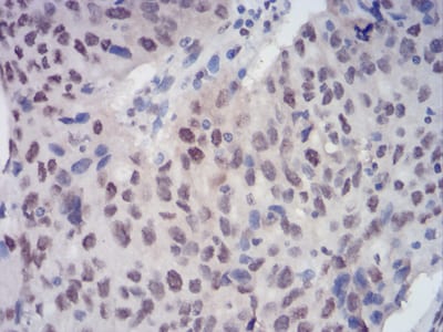
Figure 5:Immunohistochemical analysis of paraffin-embedded ovarian cancer tissues using CBX3 mouse mAb with DAB staining.
Immunohistochemical analysis

Figure 6:Immunohistochemical analysis of paraffin-embedded esophageal cancer tissues using CBX3 mouse mAb with DAB staining.
For Research Use Only. Not for use in diagnostic procedures.