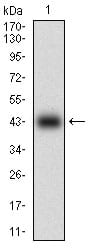
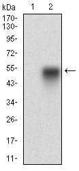
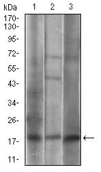
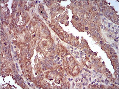
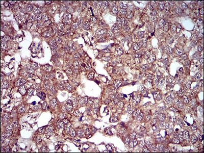
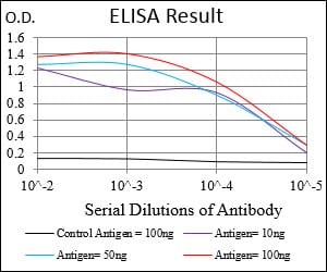
CAV2 Primary Antibody
The protein encoded by this gene is a major component of the inner surface of caveolae, small invaginations of the plasma membrane, and is involved in essential cellular functions, including signal transduction, lipid metabolism, cellular growth control and apoptosis. This protein may function as a tumor suppressor. This gene and related family member (CAV1) are located next to each other on chromosome 7, and express colocalizing proteins that form a stable hetero-oligomeric complex. Alternatively spliced transcript variants encoding different isoforms have been identified for this gene. Additional isoforms resulting from the use of alternate in-frame translation initiation codons have also been described, and shown to have preferential localization in the cell (PMID:11238462).
2. Breast Cancer Res Treat. 2008 Jul;110(2):245-56.
![Figure 1: Western blot analysis using CAV2 mAb against human CAV2 (AA: 1-86) recombinant protein. (Expected MW is 35.9 kDa)](https://cdn.shopify.com/s/files/1/0604/2661/7019/products/30645rwb_1.jpg?v=1666703186)
![Figure 2: Western blot analysis using CAV2 mAb against HEK293 (1) and CAV2 (AA: 1-86)-hIgGFc transfected HEK293 (2) cell lysate.](https://cdn.shopify.com/s/files/1/0604/2661/7019/products/30645twb_1_1b591851-db6b-40eb-bf85-a85b76df1282.jpg?v=1666703186)
![Figure 3: Western blot analysis using CAV2 mouse mAb against A549 (1), 3T3-L1 (2), A431 (3) cell lysate.](https://cdn.shopify.com/s/files/1/0604/2661/7019/products/30645lwb_1.jpg?v=1666703186)
![Figure 4: Flow cytometric analysis of A549 cells using CAV2 mouse mAb (green) and negative control (red).](https://cdn.shopify.com/s/files/1/0604/2661/7019/products/30645fcm_1_150a35d1-0eb5-47f5-9871-9a05dcc18f2b.jpg?v=1666703186)
![Figure 5: Immunohistochemical analysis of paraffin-embedded endometrial cancer tissues using CAV2 mouse mAb with DAB staining.](https://cdn.shopify.com/s/files/1/0604/2661/7019/products/30645ihc-1_1.jpg?v=1666703186)
![Figure 6: Immunohistochemical analysis of paraffin-embedded esophageal cancer tissues using CAV2 mouse mAb with DAB staining.](https://cdn.shopify.com/s/files/1/0604/2661/7019/products/30645ihc-2_1.jpg?v=1666703186)
![Black line: Control Antigen (100 ng); Purple line: Antigen(10ng); Blue line: Antigen (50 ng); Red line: Antigen (100 ng);](https://cdn.shopify.com/s/files/1/0604/2661/7019/products/30645ea_1_ee6c771a-0776-4e47-b2d3-54cccd2f7aee.jpg?v=1666703186)