

CASP-7 Primary Antibody
Item Information
Catalog #
Size
Price
Description
This gene encodes a member of the cysteine-aspartic acid protease (caspase) family. Sequential activation of caspases plays a central role in the execution-phase of cell apoptosis. Caspases exist as inactive proenzymes which undergo proteolytic processing at conserved aspartic residues to produce two subunits, large and small, that dimerize to form the active enzyme. The precursor of the encoded protein is cleaved by caspase 3 and 10, is activated upon cell death stimuli and induces apoptosis. Alternatively spliced transcript variants encoding multiple isoforms have been observed for this gene.
Product Overview
Entrez GenelD
840
Aliases
MCH3; CMH-1; LICE2; CASP7; ICE-LAP3
Clone#
4D10B2
Host / Isotype
Mouse / IgG1
Species Reactivity
Human
Immunogen
Purified recombinant fragment of human CASP-7 (AA: 29-198) expressed in E. Coli.
Formulation
Purified antibody from tissue culture in PBS with 0.05% sodium azide
Storage
Store at 4°C short term. Aliquot and store at -20°C long term. Avoid freeze/thaw cycles.
Product Applications
WB (Western Blot)
1/500 - 1/2000
IHC_P(Immunohistochemistry)
1/200 - 1/1000
FCM (Flow Cytometry)
1/200 - 1/400
ELISA
1/10000
References
1. Lung Cancer. 2009 Jul;65(1):19-24.
2. Genes Cells. 2008 Jun;13(6):609-21.
2. Genes Cells. 2008 Jun;13(6):609-21.
Product Image
Western Blot
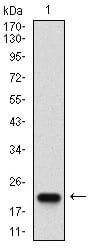
Figure 1: Western blot analysis using CASP-7 mAb against human CASP-7 (AA: 29-198) recombinant protein. (Expected MW is 22.5 kDa)
Western Blot
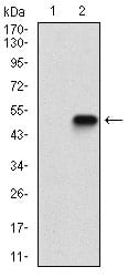
Figure 2: Western blot analysis using CASP-7 mAb against HEK293 (1) and CASP-7 (AA: 29-198)-hIgGFc transfected HEK293 (2) cell lysate.
Western Blot
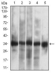
Figure 3: Western blot analysis using CASP-7 mouse mAb against Jurkat (1), HEK293 (2), MOLT4 (3), MCF-7 (4), PC-12 (5) cell lysate.
Flow cytometric
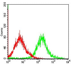
Figure 4: Flow cytometric analysis of MCF-7 cells using CASP-7 mouse mAb (green) and negative control (red).
Immunohistochemical analysis
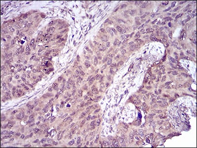
Figure 5: Immunohistochemical analysis of paraffin-embedded cervical cancer tissues using CASP-7 mouse mAb with DAB staining.
Elisa
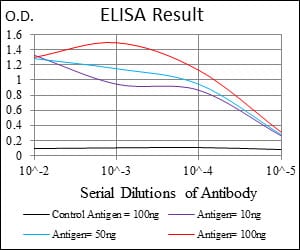
Black line: Control Antigen (100 ng); Purple line: Antigen(10ng); Blue line: Antigen (50 ng); Red line: Antigen (100 ng);
For Research Use Only. Not for use in diagnostic procedures.