

CA9 Primary Antibody
Item Information
Catalog #
Size
Price
Description
Carbonic anhydrases (CAs) are a large family of zinc metalloenzymes that catalyze the reversible hydration of carbon dioxide. They participate in a variety of biological processes, including respiration, calcification, acid-base balance, bone resorption, and the formation of aqueous humor, cerebrospinal fluid, saliva, and gastric acid. They show extensive diversity in tissue distribution and in their subcellular localization. CA IX is a transmembrane protein and the only tumor-associated carbonic anhydrase isoenzyme known. It is expressed in all clear-cell renal cell carcinoma, but is not detected in normal kidney or most other normal tissues. It may be involved in cell proliferation and transformation. This gene was mapped to 17q21.2 by fluorescence in situ hybridization, however, radiation hybrid mapping localized it to 9p13-p12.
Product Overview
Entrez GenelD
768
Aliases
MN; CAIX
Clone#
10F7A8
Host / Isotype
Mouse / IgG1
Species Reactivity
Human
Immunogen
Purified recombinant fragment of human CA9 (AA: 37-186) expressed in E. Coli.
Formulation
Purified antibody in PBS with 0.05% sodium azide
Storage
Store at 4°C short term. Aliquot and store at -20°C long term. Avoid freeze/thaw cycles.
Product Applications
WB (Western Blot)
1/500 - 1/2000
FCM (Flow Cytometry)
1/200 - 1/400
ELISA
1/10000
References
1. Breast Cancer Res Treat. 2012 Nov;136(1):67-75.
2. Histol Histopathol. 2011 Oct;26(10):1279-86.
2. Histol Histopathol. 2011 Oct;26(10):1279-86.
Product Image
Western Blot
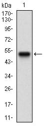
Figure 1: Western blot analysis using CA9 mAb against human CA9 recombinant protein. (Expected MW is 42 kDa)
Western Blot
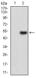
Figure 2: Western blot analysis using CA9 mAb against HEK293 (1) and CA9 (AA: 37-186)-hIgGFc transfected HEK293 (2) cell lysate.
Western Blot

Figure 3: Western blot analysis using CA9 mouse mAb against A431 (1) and SW620 (2) cell lysate.
Flow cytometric

Figure 4: Flow cytometric analysis of NTERA-2 cells using CA9 mouse mAb (green) and negative control (red).
Elisa
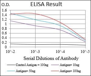
Black line: Control Antigen (100 ng); Purple line: Antigen(10ng); Blue line: Antigen (50 ng); Red line: Antigen (100 ng);
For Research Use Only. Not for use in diagnostic procedures.