

C3C Primary Antibody
Item Information
Catalog #
Size
Price
Description
Complement component C3 plays a central role in the activation of complement system. Its activation is required for both classical and alternative complement activation pathways. The encoded preproprotein is proteolytically processed to generate alpha and beta subunits that form the mature protein, which is then further processed to generate numerous peptide products. The C3a peptide, also known as the C3a anaphylatoxin, modulates inflammation and possesses antimicrobial activity. Mutations in this gene are associated with atypical hemolytic uremic syndrome and age-related macular degeneration in human patients.
Product Overview
Entrez GenelD
718
Aliases
ASP; C3a; C3b; AHUS5; ARMD9; CPAMD1; HEL-S-62p
Clone#
4C6A8
Host / Isotype
Mouse / IgG1
Species Reactivity
Human
Immunogen
Purified recombinant fragment of human C3C (AA: 1521-1649) expressed in E. Coli.
Formulation
Purified antibody in PBS with 0.05% sodium azide
Storage
Store at 4°C short term. Aliquot and store at -20°C long term. Avoid freeze/thaw cycles.
Product Applications
WB (Western Blot)
1/500 - 1/2000
IHC_P(Immunohistochemistry)
1/200 - 1/1000
ICC (Immunocytochemistry)
1/200 - 1/1000
FCM (Flow Cytometry)
1/200 - 1/400
ELISA
1/10000
References
1.Blood. 2015 Apr 9;125(15):2359-69.
2.Mediators Inflamm. 2013;2013:716902.
2.Mediators Inflamm. 2013;2013:716902.
Product Image
Elisa
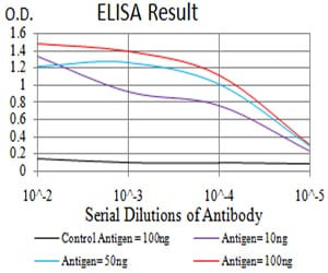
Figure 1: Black line: Control Antigen (100 ng);Purple line: Antigen (10ng); Blue line: Antigen (50 ng); Red line:Antigen (100 ng)
Western Blot
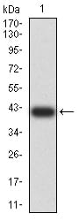
Figure 2:Western blot analysis using C3C mAb against human C3C (AA: 1521-1649) recombinant protein. (Expected MW is 40.7 kDa)
Western Blot
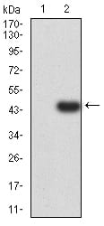
Figure 3:Western blot analysis using C3C mAb against HEK293 (1) and C3C (AA: 1521-1649)-hIgGFc transfected HEK293 (2) cell lysate.
Immunofluorescence analysis
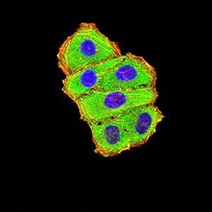
Figure 4:Immunofluorescence analysis of Hela cells using C3C mouse mAb (green). Blue: DRAQ5 fluorescent DNA dye. Red: Actin filaments have been labeled with Alexa Fluor- 555 phalloidin. Secondary antibody from Fisher (Cat#: 35503)
Flow cytometric
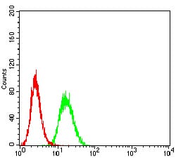
Figure 5:Flow cytometric analysis of Hela cells using C3C mouse mAb (green) and negative control (red).
Immunohistochemical analysis

Figure 6:Immunohistochemical analysis of paraffin-embedded ovarian cancer tissues using C3C mouse mAb with DAB staining.
Immunohistochemical analysis
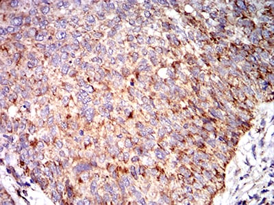
Figure 7:Immunohistochemical analysis of paraffin-embedded bladder cancer tissues using C3C mouse mAb with DAB staining.
For Research Use Only. Not for use in diagnostic procedures.