

C1QA Primary Antibody
Item Information
Catalog #
Size
Price
Description
This gene encodes a major constituent of the human complement subcomponent C1q. C1q associates with C1r and C1s in order to yield the first component of the serum complement system. Deficiency of C1q has been associated with lupus erythematosus and glomerulonephritis. C1q is composed of 18 polypeptide chains: six A-chains, six B-chains, and six C-chains. Each chain contains a collagen-like region located near the N terminus and a C-terminal globular region. The A-, B-, and C-chains are arranged in the order A-C-B on chromosome 1. This gene encodes the A-chain polypeptide of human complement subcomponent C1q.
Product Overview
Entrez GenelD
712
Aliases
N
Clone#
8B5B1
Host / Isotype
Mouse / IgG2a
Species Reactivity
Human
Immunogen
Purified recombinant fragment of human C1QA (AA: 23-167) expressed in E. Coli.
Formulation
Purified antibody in PBS with 0.05% sodium azide
Storage
Store at 4°C short term. Aliquot and store at -20°C long term. Avoid freeze/thaw cycles.
Product Applications
WB (Western Blot)
1/500 - 1/2000
IHC_P(Immunohistochemistry)
1/200 - 1/1000
FCM (Flow Cytometry)
1/200 - 1/400
ELISA
1/10000
References
1.Immunology. 2012 May;136(1):78-85.
2.Br J Cancer. 2010 Apr 13;102(8):1294-9.
2.Br J Cancer. 2010 Apr 13;102(8):1294-9.
Product Image
Elisa
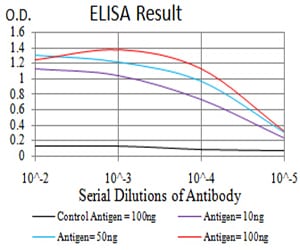
Figure 1: Black line: Control Antigen (100 ng);Purple line: Antigen (10ng); Blue line: Antigen (50 ng); Red line:Antigen (100 ng)
Western Blot
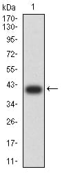
Figure 2:Western blot analysis using C1QA mAb against human C1QA (AA: 23-167) recombinant protein. (Expected MW is 40.6 kDa)
Western Blot
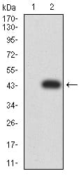
Figure 3:Western blot analysis using C1QA mAb against HEK293 (1) and C1QA (AA: 23-167)-hIgGFc transfected HEK293 (2) cell lysate.
Flow cytometric
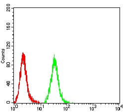
Figure 4:Flow cytometric analysis of Hela cells using C1QA mouse mAb (green) and negative control (red).
Immunohistochemical analysis
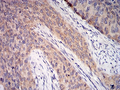
Figure 5:Immunohistochemical analysis of paraffin-embedded cervical cancer tissues using C1QA mouse mAb with DAB staining.
Immunohistochemical analysis
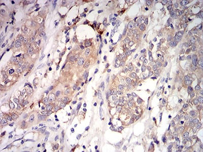
Figure 6:Immunohistochemical analysis of paraffin-embedded bladder cancer tissues using C1QA mouse mAb with DAB staining.
For Research Use Only. Not for use in diagnostic procedures.