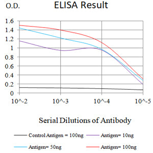
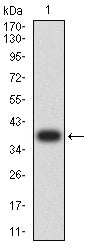
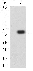
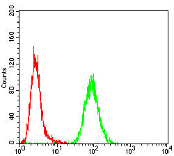
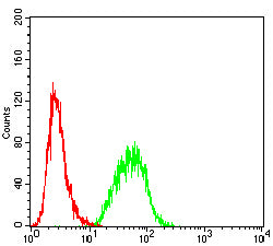
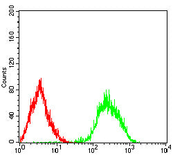
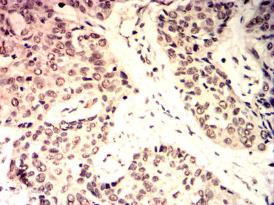
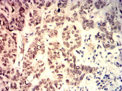
C-MYC Primary Antibody

Item Information
Catalog #
Size
Price
Description

This gene is a proto-oncogene and encodes a nuclear phosphoprotein that plays a role in cell cycle progression, apoptosis and cellular transformation. The encoded protein forms a heterodimer with the related transcription factor MAX. This complex binds to the E box DNA consensus sequence and regulates the transcription of specific target genes. Amplification of this gene is frequently observed in numerous human cancers. Translocations involving this gene are associated with Burkitt lymphoma and multiple myeloma in human patients. There is evidence to show that translation initiates both from an upstream, in-frame non-AUG (CUG) and a downstream AUG start site, resulting in the production of two isoforms with distinct N-termini. [provided by RefSeq, Aug 2017]

Product Overview
Entrez GenelD
4609
Aliases
MRTL; MYCC; c-Myc; bHLHe39
Clone#
6H11F6
Host / Isotype
Mouse / Mouse IgG1
Immunogen
Purified recombinant fragment of human C-MYC (AA: (290-439)) expressed in E. Coli.
Formulation
Purified antibody in PBS with 0.05% sodium azide
Storage
4°C; -20°C for long term storage
Product Applications
WB (Western Blot)
1/500 - 1/2000
IHC_P(Immunohistochemistry)
1/200-1/1000
FCM (Flow Cytometry)
1/200-1/400
ELISA
1/10000
References
1,Jpn J Clin Oncol. 2020 Apr 7;50(4):446-455. 2,Proc Natl Acad Sci U S A. 2020 Mar 10;117(10):5269-5279.
Product Image
ELISA
Figure 1: Black line: Control Antigen (100 ng);Purple line: Antigen (10ng); Blue line: Antigen (50 ng); Red line: Antigen (100 ng)
WESTERN BLOT
Figure 2: Western blot analysis using C-MYC mAb against human C-MYC (AA: (290-439)) recombinant protein. (Expected MW is *** kDa)
WESTERN BLOT
Figure 3: Western blot analysis using C-MYC mAb against HEK293-6e (1) and C-MYC (AA: (290-439))-hIgGFc transfected HEK293-6e (2) cell lysate.
FLOW CYTOMETRY
Figure 4: Flow cytometric analysis of Hela cells using C-MYC mouse mAb (green) and negative control (red).
FLOW CYTOMETRY
Figure 5: Flow cytometric analysis of Hepg2 cells using C-MYC mouse mAb (green) and negative control (red).
FLOW CYTOMETRY
Figure 6: Flow cytometric analysis of Lovo cells using C-MYC mouse mAb (green) and negative control (red).
IMMUNOHISTOCHEMISTRY
Figure 7: Immunohistochemical analysis of paraffin-embedded esophageal cancer tissues using C-MYC mouse mAb with DAB staining.
IMMUNOHISTOCHEMISTRY
Figure 8: Immunohistochemical analysis of paraffin-embedded lung cancer tissues using C-MYC mouse mAb with DAB staining.
For Research Use Only. Not for use in diagnostic procedures.