

BTRC Primary Antibody
Item Information
Catalog #
Size
Price
Description
This gene encodes a member of the F-box protein family which is characterized by an approximately 40 amino acid motif, the F-box. The F-box proteins constitute one of the four subunits of ubiquitin protein ligase complex called SCFs (SKP1-cullin-F-box), which function in phosphorylation-dependent ubiquitination. The F-box proteins are divided into 3 classes: Fbws containing WD-40 domains, Fbls containing leucine-rich repeats, and Fbxs containing either different protein-protein interaction modules or no recognizable motifs. The protein encoded by this gene belongs to the Fbws class; in addition to an F-box, this protein contains multiple WD-40 repeats. The encoded protein mediates degradation of CD4 via its interaction with HIV-1 Vpu. It has also been shown to ubiquitinate phosphorylated NFKBIA (nuclear factor of kappa light polypeptide gene enhancer in B-cells inhibitor, alpha), targeting it for degradation and thus activating nuclear factor kappa-B. Alternatively spliced transcript variants have been described. A related pseudogene exists in chromosome 6.
Product Overview
Entrez GenelD
8945
Aliases
FWD1; FBW1A; FBXW1; bTrCP; FBXW1A; bTrCP1; betaTrCP; BETA-TRCP
Clone#
3D5E6
Host / Isotype
Mouse / IgG1
Species Reactivity
Human
Immunogen
Purified recombinant fragment of human BTRC (AA: 24-151) expressed in E. Coli.
Formulation
Purified antibody in PBS with 0.05% sodium azide
Storage
Store at 4°C short term. Aliquot and store at -20°C long term. Avoid freeze/thaw cycles.
Product Applications
WB (Western Blot)
1/500 - 1/2000
IHC_P(Immunohistochemistry)
1/200 - 1/1000
ELISA
1/10000
References
1.Biochem Biophys Res Commun. 2013 Nov 29;441(4):831-7.
2.PLoS One. 2011;6(11):e27464.
2.PLoS One. 2011;6(11):e27464.
Product Image
Elisa

Figure 1: Black line: Control Antigen (100 ng);Purple line: Antigen (10ng); Blue line: Antigen (50 ng); Red line:Antigen (100 ng)
Western Blot
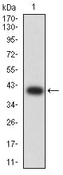
Figure 2:Western blot analysis using BTRC mAb against human BTRC (AA: 24-151) recombinant protein. (Expected MW is 40.2 kDa)
Western Blot

Figure 3:Western blot analysis using BTRC mAb against HEK293 (1) and BTRC (AA: 24-151)-hIgGFc transfected HEK293 (2) cell lysate.
Western Blot

Figure 4:Western blot analysis using BTRC mouse mAb against Ramos (1), MCF-7 (2), and K562 (3) cell lysate.
Immunohistochemical analysis
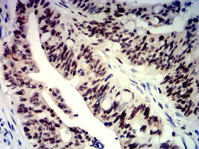
Figure 5:Immunohistochemical analysis of paraffin-embedded rectum cancer tissues using BTRC mouse mAb with DAB staining.
For Research Use Only. Not for use in diagnostic procedures.