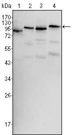
BRAF Primary Antibody

Item Information
Catalog #
Size
Price
Description

BRAF: v-raf murine sarcoma viral oncogene homolog B1, also known as BRAF1; RAFB1; B-RAF1; FLJ95109. Entrez Protein NP_004324. It is the main effectors recruited by GTP-bound Ras to activate the MEK-MAP kinase pathway. B-Raf contains three consensus Akt phosphorylationsites (Ser364, Ser428, and Thr439). B-Raf is a key regulatory molecule of the mitogen-activated protein kinase kinase (MEK), it has a long amino-terminal region,the region is essential for homo-dimerization of B-Raf and hetero-dimerization of B-Raf and c-Raf at the plasma membrane, followed by phosphorylation of Thr118 in the amino-terminal B-Raf-specific region. Notably, in calcium ionophore-stimulated HeLa cells, B-Raf could propagate signals to MEK under the basal level of GTP-Ras. Expression of Raf-B is highly restricted with highestlevels in the cerebrum and testes and defects in braf are involved in a wide range of cancers. The BRAF gene mutation is frequently detected in papillary thyroid carcinoma,melanocytic nevi, primary cutaneous melanomas and colorectal cancers.

Product Overview
Entrez GenelD
673
Aliases
BRAF1; RAFB1; B-RAF1; FLJ95109
Clone#
1H12
Host / Isotype
Mouse / IgG1
Species Reactivity
Human, Mouse
Immunogen
Purified recombinant fragment of human BRAF expressed in E. Coli.
Formulation
Ascitic fluid containing 0.03% sodium azide.
Storage
4°C; -20°C for long term storage
Product Applications
WB (Western Blot)
1/500 - 1/2000
IHC_P(Immunohistochemistry)
1/200 - 1/1000
ICC (Immunocytochemistry)
1/200 - 1/1000
ELISA
1/10000
References
1. J Clin Oncol. 2008 Dec 10;26(35):5705-12.
2. Endocr Relat Cancer. 2006 Mar;13(1):257-69.
Product Image
Western Blot
Figure 1: Western blot analysis using BRAF mouse mAb against Hela (1), HL60 (2), HepG2 (3) and NIH/3T3 (4) cell lysate.
Immunohistochemical analysis
Figure 2: Immunohistochemical analysis of paraffin-embedded human testis tissues using BRAF mouse mAb.
Immunofluorescence analysis
Figure 3: Confocal Immunofluorescence analysis of MCF-7 (left) and HepG2 (right) cells using BRAF mouse mAb (green). Red: Actin filaments have been labeled with DY-554 phalloidin. Blue: DRAQ5 fluorescent DNA dye
For Research Use Only. Not for use in diagnostic procedures.