
BNIP3 Primary Antibody
Item Information
Catalog #
Size
Price
Description
This gene is encodes a mitochondrial protein that contains a BH3 domain and acts as a pro-apoptotic factor. The encoded protein interacts with anti-apoptotic proteins, including the E1B 19 kDa protein and Bcl2. This gene is silenced in tumors by DNA methylation.
Product Overview
Entrez GenelD
664
Aliases
NIP3
Clone#
6A5F7
Host / Isotype
Mouse / IgG2a
Species Reactivity
Human
Immunogen
Purified recombinant fragment of human BNIP3 (AA: 50-155) expressed in E. Coli.
Formulation
Purified antibody in PBS with 0.05% sodium azide
Storage
Store at 4°C short term. Aliquot and store at -20°C long term. Avoid freeze/thaw cycles.
Product Applications
WB (Western Blot)
1/500 - 1/2000
IHC_P(Immunohistochemistry)
1/200 - 1/1000
ICC (Immunocytochemistry)
1/50 - 1/250
FCM (Flow Cytometry)
1/200 - 1/400
ELISA
1/10000
References
1.Tumour Biol. 2015 Jun;36(6):4731-40.
2.PLoS One. 2014 May 13;9(5):e96733.
2.PLoS One. 2014 May 13;9(5):e96733.
Product Image
Elisa
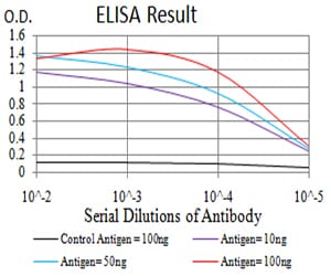
Figure 1: Black line: Control Antigen (100 ng);Purple line: Antigen (10ng); Blue line: Antigen (50 ng); Red line:Antigen (100 ng)
Western Blot
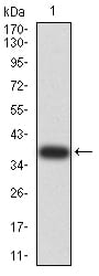
Figure 2:Western blot analysis using BNIP3 mAb against human BNIP3 (AA: 50-155) recombinant protein. (Expected MW is 38 kDa)
Western Blot
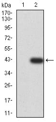
Figure 3:Western blot analysis using BNIP3 mAb against HEK293 (1) and BNIP3 (AA: 50-155)-hIgGFc transfected HEK293 (2) cell lysate.
Immunofluorescence analysis
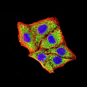
Figure 4:Immunofluorescence analysis of Hela cells using BNIP3 mouse mAb (green). Blue: DRAQ5 fluorescent DNA dye. Red: Actin filaments have been labeled with Alexa Fluor- 555 phalloidin. Secondary antibody from Fisher (Cat#: 35503)
Flow cytometric
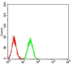
Figure 5:Flow cytometric analysis of Hela cells using BNIP3 mouse mAb (green) and negative control (red).
Immunohistochemical analysis
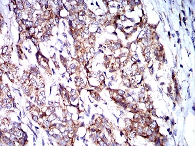
Figure 6:Immunohistochemical analysis of paraffin-embedded breast cancer tissues using BNIP3 mouse mAb with DAB staining.
Immunohistochemical analysis
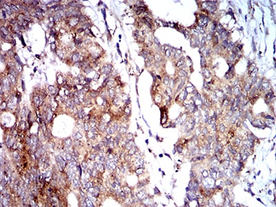
Figure 7:Immunohistochemical analysis of paraffin-embedded rectum cancer tissues using BNIP3 mouse mAb with DAB staining.
For Research Use Only. Not for use in diagnostic procedures.