
BMP7 Primary Antibody
Item Information
Catalog #
Size
Price
Description
The bone morphogenetic proteins (BMPs) are a family of secreted signaling molecules that can induce ectopic bone growth. Many BMPs are part of the transforming growth factor-beta (TGFB) superfamily. BMPs were originally identified by an ability of demineralized bone extract to induce endochondral osteogenesis in vivo in an extraskeletal site. Based on its expression early in embryogenesis, the BMP encoded by this gene has a proposed role in early development and possible bone inductive activity.
Product Overview
Entrez GenelD
655
Aliases
OP-1
Clone#
6E5D12
Host / Isotype
Mouse / IgG1
Species Reactivity
Human, Mouse
Immunogen
Purified recombinant fragment of human BMP7 (AA: 239-431) expressed in E. Coli.
Formulation
Purified antibody in PBS with 0.05% sodium azide
Storage
Store at 4°C short term. Aliquot and store at -20°C long term. Avoid freeze/thaw cycles.
Product Applications
WB (Western Blot)
1/500 - 1/2000
IHC_P(Immunohistochemistry)
1/200 - 1/1000
FCM (Flow Cytometry)
1/200 - 1/400
ELISA
1/10000
References
1.Immunol Cell Biol. 2014 May-Jun;92(5):427-35.
2.J Exp Med. 2013 Nov 18;210(12):2597-610.
2.J Exp Med. 2013 Nov 18;210(12):2597-610.
Product Image
Elisa
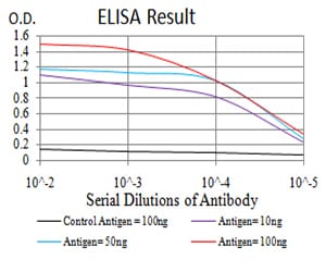
Figure 1: Black line: Control Antigen (100 ng); Purple line: Antigen(10ng); Blue line: Antigen (50 ng); Red line: Antigen (100 ng);
Western Blot

Figure 2:Western blot analysis using BMP7 mAb against human BMP7 (AA: 239-431) recombinant protein. (Expected MW is 47.7 kDa)
Western Blot

Figure 3:Western blot analysis using BMP7 mAb against HEK293 (1) and BMP7 (AA: 239-431)-hIgGFc transfected HEK293 (2) cell lysate.
Western Blot

Figure 4:Western blot analysis using BMP7 mouse mAb against Raw264.7 (1), A549 (2), Jurkat (3), PC-3 (4), HEK293 (5), Jurkat (6), NIH/3T3 (7), and Hela (8) cell lysate.
Flow cytometric
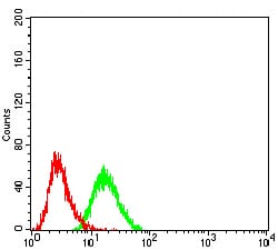
Figure 5:Flow cytometric analysis of HEK293 cells using BMP7 mouse mAb (green) and negative control (red).
Immunohistochemical analysis

Figure 6:Immunohistochemical analysis of paraffin-embedded esophageal cancer tissues using BMP7 mouse mAb with DAB staining.
For Research Use Only. Not for use in diagnostic procedures.