

BIN1 Primary Antibody
Item Information
Catalog #
Size
Price
Description
This gene encodes several isoforms of a nucleocytoplasmic adaptor protein, one of which was initially identified as a MYC-interacting protein with features of a tumor suppressor. Isoforms that are expressed in the central nervous system may be involved in synaptic vesicle endocytosis and may interact with dynamin, synaptojanin, endophilin, and clathrin. Isoforms that are expressed in muscle and ubiquitously expressed isoforms localize to the cytoplasm and nucleus and activate a caspase-independent apoptotic process. Studies in mouse suggest that this gene plays an important role in cardiac muscle development. Alternate splicing of the gene results in several transcript variants encoding different isoforms. Aberrant splice variants expressed in tumor cell lines have also been described.
Product Overview
Entrez GenelD
274
Aliases
AMPH2; AMPHL; SH3P9
Clone#
3B6A4
Host / Isotype
Mouse / IgG2b
Species Reactivity
Human, Mouse
Immunogen
Purified recombinant fragment of human BIN1 (AA: 189-398) expressed in E. Coli.
Formulation
Purified antibody in PBS with 0.05% sodium azide
Storage
Store at 4°C short term. Aliquot and store at -20°C long term. Avoid freeze/thaw cycles.
Product Applications
WB (Western Blot)
1/500 - 1/2000
FCM (Flow Cytometry)
1/200 - 1/400
ELISA
1/10000
References
1.Trends Mol Med. 2013 Oct;19(10):594-603.
2.Mol Med. 2012 May 9;18:507-18.
2.Mol Med. 2012 May 9;18:507-18.
Product Image
Elisa
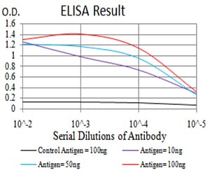
Figure 1: Black line: Control Antigen (100 ng);Purple line: Antigen (10ng); Blue line: Antigen (50 ng); Red line:Antigen (100 ng)
Western Blot
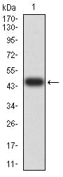
Figure 2:Western blot analysis using BIN1 mAb against human BIN1 (AA: 189-398) recombinant protein. (Expected MW is 47.1 kDa)
Western Blot
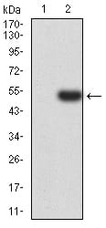
Figure 3:Western blot analysis using BIN1 mAb against HEK293 (1) and BIN1 (AA: 189-398)-hIgGFc transfected HEK293 (2) cell lysate.
Western Blot
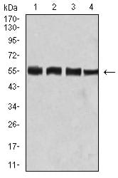
Figure 4:Western blot analysis using BIN1 mouse mAb against Hela (1), C2C12 (2), A431 (3), and HEK293 (4) cell lysate.
Flow cytometric
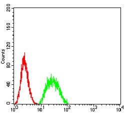
Figure 5:Flow cytometric analysis of Hela cells using BIN1 mouse mAb (green) and negative control (red).
For Research Use Only. Not for use in diagnostic procedures.