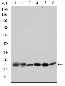
BID Primary Antibody

Item Information
Catalog #
Size
Price
Description

This gene encodes a death agonist that heterodimerizes with either agonist BAX or antagonist BCL2. The encoded protein is a member of the BCL-2 family of cell death regulators. It is a mediator of mitochondrial damage induced by caspase-8 (CASP8); CASP8 cleaves this encoded protein, and the COOH-terminal part translocates to mitochondria where it triggers cytochrome c release. Multiple alternatively spliced transcript variants have been found, but the full-length nature of some variants has not been defined.Tissue specificity: Isoform 2 and isoform 3 are expressed in spleen, bone marrow, cerebral and cerebellar cortex. Isoform 2 is expressed in spleen, pancreas and placenta (at protein level). Isoform 3 is expressed in lung, pancreas and spleen (at protein level). Isoform 4 is expressed in lung and pancreas (at protein level)

Product Overview
Entrez GenelD
637
Aliases
FP497; MGC15319; MGC42355; BID
Clone#
3C5
Host / Isotype
Mouse / IgG1
Species Reactivity
Human
Immunogen
Purified recombinant full length protein of human BID expressed in E. Coli
Formulation
Ascitic fluid containing 0.03% sodium azide.
Storage
4°C; -20°C for long term storage
Product Applications
WB (Western Blot)
1/500 - 1/2000
IHC_P(Immunohistochemistry)
1/200 - 1/1000
ICC (Immunocytochemistry)
1/200 - 1/1000
FCM (Flow Cytometry)
1/200 - 1/400
ELISA
1/10000
References
1. Photochem Photobiol. 2008 Jan-Feb;84(1):250-7.
2. Cell Signal. 2007 Dec;19(12):2468-78.
Product Image
Western Blot
Figure 1: Western blot analysis using BID mouse mAb against Hela (1), A431 (2), Jurkat (3), A549 (4), HepG2 (5), and HEK293 (6) cell lysate.
Immunohistochemical analysis
Figure 2: Immunohistochemical analysis of paraffin-embedded prostate tissues (left) and tonsil tissues (right) using BID mouse mAb with DAB staining.
Immunofluorescence analysis
Figure 3: Immunofluorescence analysis of Hela cells using BID mouse mAb (green). Blue: DRAQ5 fluorescent DNA dye. Red: Actin filaments have been labeled with Alexa Fluor-555 phalloidin.
Flow cytometric
Figure 4: Flow cytometric analysis of Hela cells using BID mouse mAb (green) and negative control (purple).
Elisa
Red: Control Antigen (100ng); Purple: Antigen (10ng); Green: Antigen (50ng); Blue: Antigen (100ng);
For Research Use Only. Not for use in diagnostic procedures.