

BCL2L2 Primary Antibody
Item Information
Catalog #
Size
Price
Description
This gene encodes a member of the BCL-2 protein family. The proteins of this family form hetero- or homodimers and act as anti- and pro-apoptotic regulators. Expression of this gene in cells has been shown to contribute to reduced cell apoptosis under cytotoxic conditions. Studies of the related gene in mice indicated a role in the survival of NGF- and BDNF-dependent neurons. Mutation and knockout studies of the mouse gene demonstrated an essential role in adult spermatogenesis. Alternative splicing results in multiple transcript variants. Read-through transcription also exists between this gene and the neighboring downstream PABPN1 (poly(A) binding protein, nuclear 1) gene.
Product Overview
Entrez GenelD
599
Aliases
BCLW; BCL-W; PPP1R51; BCL2-L-2
Clone#
4G12E6
Host / Isotype
Mouse / IgG2a
Species Reactivity
Human
Immunogen
Purified recombinant fragment of human BCL2L2 (AA: 6-118) expressed in E. Coli.
Formulation
Purified antibody in PBS with 0.05% sodium azide
Storage
Store at 4°C short term. Aliquot and store at -20°C long term. Avoid freeze/thaw cycles.
Product Applications
WB (Western Blot)
1/500 - 1/2000
IHC_P(Immunohistochemistry)
1/200 - 1/1000
ICC (Immunocytochemistry)
1/200 - 1/1000
FCM (Flow Cytometry)
1/200 - 1/400
ELISA
1/10000
References
1.Med Sci Monit. 2016 Nov 1;22:4139-4145.
2.Food Chem Toxicol. 2010 Aug-Sep;48(8-9):2259-64.
2.Food Chem Toxicol. 2010 Aug-Sep;48(8-9):2259-64.
Product Image
Elisa

Figure 1: Black line: Control Antigen (100 ng);Purple line: Antigen (10ng); Blue line: Antigen (50 ng); Red line:Antigen (100 ng)
Western Blot

Figure 2:Western blot analysis using BCL2L2 mAb against human BCL2L2 (AA: 6-118) recombinant protein. (Expected MW is 38.2 kDa)
Western Blot

Figure 3:Western blot analysis using BCL2L2 mAb against HEK293 (1) and BCL2L2 (AA: 6-118)-hIgGFc transfected HEK293 (2) cell lysate.
Western Blot

Figure 4:Western blot analysis using BCL2L2 mouse mAb against HCT116 (1), LOVO (2), SW480 (3), and HL-60 (4) cell lysate.
Immunofluorescence analysis
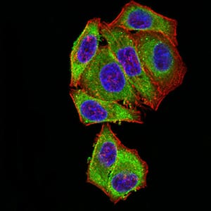
Figure 5:Immunofluorescence analysis of Hela cells using BCL2L2 mouse mAb (green). Blue: DRAQ5 fluorescent DNA dye. Red: Actin filaments have been labeled with Alexa Fluor- 555 phalloidin. Secondary antibody from Fisher (Cat#: 35503)
Flow cytometric

Figure 6:Flow cytometric analysis of Hela cells using BCL2L2 mouse mAb (green) and negative control (red).
Flow cytometric

Figure 7:Flow cytometric analysis of K562 cells using BCL2L2 mouse mAb (green) and negative control (red).
Immunohistochemical analysis

Figure 8:Immunohistochemical analysis of paraffin-embedded cervical cancer tissues using BCL2L2 mouse mAb with DAB staining.
Immunohistochemical analysis

Figure 9:Immunohistochemical analysis of paraffin-embedded ovarian cancer tissues using BCL2L2 mouse mAb with DAB staining.
For Research Use Only. Not for use in diagnostic procedures.