
BCL-10 Primary Antibody
Item Information
Catalog #
Size
Price
Description
Bcl-10 (B-cell CLL/lymphoma 10), also known as CLAP, Me10, CIPER, c-E10, CARMEN. Entrez Protein NP_003912. It is a protein containing a caspase recruitment domain (CARD). It plays an important
role in apoptosis and activating NF-kappaB. The research suggested that it interacted with other
CARD domain containing proteins including CARD9, 10, 11 and 14, which were thought to function as
upstream regulators in NF-kappaB signaling. Bcl-10 is found to form a complex with MALT1 which
encoded by another gene known to be translocated in MALT lymphoma. MALT1 and Bcl-10 are thought to
synergize in the activation of NF-kappaB, and the deregulation of either of them may contribute to
the same pathogenetic process that leads to the malignancy.
Product Overview
Entrez GenelD
8915
Aliases
CLAP; Me10; CIPER; c-E10; CARMEN
Clone#
4F8
Host / Isotype
Mouse / IgG1
Species Reactivity
Human, Mouse
Immunogen
Purified recombinant fragment of human BCL-10 expressed in E. Coli.
Formulation
Ascitic fluid containing 0.03% sodium azide.
Storage
Store at 4°C short term. Aliquot and store at -20°C long term. Avoid freeze/thaw cycles.
Product Applications
WB (Western Blot)
1/500 - 1/2000
IHC_P(Immunohistochemistry)
1/200 - 1/1000
ICC (Immunocytochemistry)
1/200 - 1/1000
FCM (Flow Cytometry)
1/200 - 1/400
ELISA
1/10000
References
1. Willis, T.G., et al. (1999) Cell. 96, 35-45.
2. Lucas, P.C., et al. (2001) J. Biol.Chem. 276, 19012-19019.
3. Wang, L., et al. (2001) J. Biol.Chem. 276, 21405-21409
2. Lucas, P.C., et al. (2001) J. Biol.Chem. 276, 19012-19019.
3. Wang, L., et al. (2001) J. Biol.Chem. 276, 21405-21409
Product Image
Western Blot

Figure 1: Western blot analysis using BCL10 mouse mAb against NIH/3T3 (1), Hela (2), MCF-7 (3) and Jurkat (4) cell lysate.
Immunohistochemical analysis

Figure 2: Immunohistochemical analysis of paraffin-embedded human breast carcinoma (A) and liver carcinoma (B), showing cytoplasmic localization using BCL10 mouse mAb with DAB staining.
Immunofluorescence analysis

Figure 3: Confocal Immunofluorescence analysis of Hela cells using BCL10 mouse mAb (green). Red: Actin filaments have been labeled with Alexa Fluor-555 phalloidin. Blue: DRAQ5 fluorescent DNA dye.
Flow cytometric
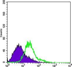
Figure 4: Flow cytometric analysis of Hela cells using BCL10 mouse mAb (green) and negative control (purple).
For Research Use Only. Not for use in diagnostic procedures.