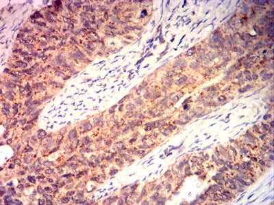
BAX Primary Antibody

Item Information
Catalog #
Size
Price
Description

The protein encoded by this gene belongs to the BCL2 protein family. BCL2 family members form hetero- or homodimers and act as anti- or pro-apoptotic regulators that are involved in a wide variety of cellular activities. This protein forms a heterodimer with BCL2, and functions as an apoptotic activator. The association and the ratio of BAX to BCL2 also determines survival or death of a cell following an apoptotic stimulus. This protein is reported to interact with, and increase the opening of, the mitochondrial voltage-dependent anion channel (VDAC), which leads to the loss in membrane potential and the release of cytochrome c. The expression of this gene is regulated by the tumor suppressor P53 and has been shown to be involved in P53-mediated apoptosis. Multiple alternatively spliced transcript variants, which encode different isoforms, have been reported for this gene. [provided by RefSeq, Dec 2019]

Product Overview
Entrez GenelD
581
Aliases
BCL2L4
Clone#
7D8B2
Host / Isotype
Mouse / Mouse IgG2b
Immunogen
Purified recombinant fragment of human BAX (AA:(13-160)) expressed in E. Coli.
Formulation
Purified antibody in PBS with 0.05% sodium azide
Storage
4°C; -20°C for long term storage
Product Applications
WB (Western Blot)
1/500 - 1/2000
IHC_P(Immunohistochemistry)
1/200-1/1000
FCM (Flow Cytometry)
1/200-1/400
ELISA
1/10000
References
1,J Biol Chem. 2019 Dec 13;294(50):19055-19065.2,Biochem Biophys Res Commun. 2019 Jun 30;514(3):881-887.
Product Image
ELISA
Figure 1: Black line: Control Antigen (100 ng);Purple line: Antigen (10ng); Blue line: Antigen (50 ng); Red line: Antigen (100 ng)
WESTERN BLOT
Figure 2: Western blot analysis using BAX mAb against human BAX (AA:(13-160)) recombinant protein. (Expected MW is 36.3 kDa)
WESTERN BLOT
Figure 3: Western blot analysis using BAX mouse mAb against Hela (1), C2C12 (2),C6 (3),HepG2 (4),MCF-7 (5),Ramos (6),Raji (7),HEK293 (8), and HEK293-6e (9) cell lysate.
FLOW CYTOMETRY
Figure 4: Flow cytometric analysis of Hela cells using BAX mouse mAb (green) and negative control (red).
FLOW CYTOMETRY
Figure 5: Flow cytometric analysis of Hepg2 cells using BAX mouse mAb (green) and negative control (red).
FLOW CYTOMETRY
Figure 6: Flow cytometric analysis of Jurkat cells using BAX mouse mAb (green) and negative control (red).
IMMUNOHISTOCHEMISTRY
Figure 7: Immunohistochemical analysis of paraffin-embedded cervical cancer tissues using BAX mouse mAb with DAB staining.
IMMUNOHISTOCHEMISTRY
Figure 8: Immunohistochemical analysis of paraffin-embedded esophageal cancer tissues using BAX mouse mAb with DAB staining.
For Research Use Only. Not for use in diagnostic procedures.